p.6 抽出テキスト
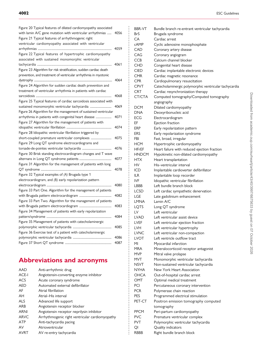
Figure 20 Typical features of dilated cardiomyopathy associated with lamin A/C gene mutation with ventricular arrhythmias ...... 4056 Figure 21 Typical features of arrhythmogenic right ventricular cardiomyopathy associated with ventricular arrhythmias ................................................................................................ 4059 Figure 22 Typical features of hypertrophic cardiomyopathy associated with sustained monomorphic ventricular tachycardia ................................................................................................. 4061 Figure 23 Algorithm for risk stratification, sudden cardiac death prevention, and treatment of ventricular arrhythmia in myotonic dystrophy .......................................................................................................................... 4064 Figure 24 Algorithm for sudden cardiac death prevention and treatment of ventricular arrhythmia in patients with cardiac sarcoidosis .......................................................................................................... 4068 Figure 25 Typical features of cardiac sarcoidosis associated with sustained monomorphic ventricular tachycardia .............................. 4069 Figure 26 Algorithm for the management of sustained ventricular arrhythmia in patients with congenital heart disease ...................... 4071 Figure 27 Algorithm for the management of patients with idiopathic ventricular fibrillation ............................................................... 4074 Figure 28 Idiopathic ventricular fibrillation triggered by short-coupled premature ventricular complexes ............................. 4075 Figure 29 Long QT syndrome electrocardiograms and torsade-de-pointes ventricular tachycardia ......................................... 4076 Figure 30 Brisk standing electrocardiogram changes and T wave alternans in Long QT syndrome patients ............................................. 4077 Figure 31 Algorithm for the management of patients with long QT syndrome ................................................................................................... 4078 Figure 32 Typical examples of (A) Brugada type 1 electrocardiogram, and (B) early repolarization pattern electrocardiogram ........................................................................................... 4080 Figure 33 Part One. Algorithm for the management of patients with Brugada pattern electrocardiogram ............................................. 4082 Figure 33 Part Two. Algorithm for the management of patients with Brugada pattern electrocardiogram ............................................. 4083 Figure 34 Management of patients with early repolarization pattern/syndrome ........................................................................................... 4084 Figure 35 Management of patients with catecholaminergic polymorphic ventricular tachycardia ...................................................... 4085 Figure 36 Exercise test of a patient with catecholaminergic polymorphic ventricular tachycardia ...................................................... 4086 Figure 37 Short QT syndrome ................................................................. 4087

Abbreviations and acronyms

AAD Anti-arrhythmic drug ACE-I Angiotensin-converting enzyme inhibitor ACS Acute coronary syndrome AED Automated external defibrillator AF Atrial fibrillation AH Atrial–His interval ALS Advanced life support ARB Angiotensin receptor blocker ARNI Angiotensin receptor neprilysin inhibitor ARVC Arrhythmogenic right ventricular cardiomyopathy ATP Anti-tachycardia pacing AV Atrioventricular AVRT AV re-entry tachycardia BBR-VT Bundle branch re-entrant ventricular tachycardia BrS Brugada syndrome CA Cardiac arrest cAMP Cyclic adenosine monophosphate CAD Coronary artery disease CAG Coronary angiogram CCB Calcium channel blocker CHD Congenital heart disease CIED Cardiac implantable electronic devices CMR Cardiac magnetic resonance CPR Cardiopulmonary resuscitation CPVT Catecholaminergic polymorphic ventricular tachycardia CRT Cardiac resynchronization therapy CT/CTA Computed tomography/Computed tomography angiography DCM Dilated cardiomyopathy DNA Deoxyribonucleic acid ECG Electrocardiogram EF Ejection fraction ERP Early repolarization pattern ERS Early repolarization syndrome FBI Fast, broad, irregular HCM Hypertrophic cardiomyopathy HFrEF Heart failure with reduced ejection fraction HNDCM Hypokinetic non-dilated cardiomyopathy HTX Heart transplantation HV His–ventricular interval ICD Implantable cardioverter defibrillator ILR Implantable loop recorder IVF Idiopathic ventricular fibrillation LBBB Left bundle branch block LCSD Left cardiac sympathetic denervation LGE Late gadolinium enhancement LMNA Lamin A/C LQTS Long QT syndrome LV Left ventricular LVAD Left ventricular assist device LVEF Left ventricular ejection fraction LVH Left ventricular hypertrophy LVNC Left ventricular non-compaction LVOT Left ventricle outflow tract MI Myocardial infarction MRA Mineralocorticoid receptor antagonist MVP Mitral valve prolapse MVT Monomorphic ventricular tachycardia NSVT Non-sustained ventricular tachycardia NYHA New York Heart Association OHCA Out-of-hospital cardiac arrest OMT Optimal medical treatment PCI Percutaneous coronary intervention PCR Polymerase chain reaction PES Programmed electrical stimulation PET-CT Positron emission tomography computed tomography PPCM Peri-partum cardiomyopathy PVC Premature ventricular complex PVT Polymorphic ventricular tachycardia QI Quality indicators RBBB Right bundle branch block Downloaded from academic.oup.com/eurheartj/article/43/40/3997/6675633 by guest on 07 October 2026