p.56 抽出テキスト
7.1.2.2 Premature ventricular complex-induced/-aggravated

cardiomyopathy The importance of PVC-induced cardiomyopathy as a secondary and reversible cause of LV dysfunction in patients without SHD has been recognized.607,608 The patient’s medical and family history, 12-lead ECG, Holter-ECG, and echocardiography form the cornerstones of the evaluation of patients with suspected PVC-induced cardiomyopathy (Figure 18). PVC burden has been shown to be the strongest independent predictor of PVC-induced cardiomyopathy in several studies.600,609–611 Day-by-day fluctuations of PVC burden have been reported in patients undergoing 14-day monitoring, but most data are based on 24-h registrations.611 A PVC burden of at least 10% appears to be the minimal threshold for development of PVC-induced cardiomyopathy, and the risk increases further with a PVC burden .20%.600,611 In patients with a PVC burden ,10%, other cardiomyopathy aetiologies should be suspected and further diagnostic work-up undertaken. In such patients, Holter-ECG should be repeated to assess fluctuations in PVC burden. Factors predicting adverse LV remodelling in patients with frequent PVCs include superior PVC axis, epicardial origin, NSVT, shorter coupling interval, and male gender.535,611–613 Frequent PVCs can also aggravate LV dysfunction in patients with SHD. In such cases, LV dysfunction can either be a direct consequence of PVCs as in PVC-induced cardiomyopathy, or due to the limiting effect of PVCs on optimal biventricular pacing in CRT patients. Parameters such as smaller LV end diastolic diameter and shorter intrinsic QRS duration might help to distinguish PVC-induced cardiomyopathy from PVC-aggravated cardiomyopathy.614 CMR should be considered for patients suspected to have PVC-induced cardiomyopathy to exclude subtle forms of SHD.590,615 In a patient with frequent PVCs, the presence of LGE suggests SHD with frequent PVCs rather than PVC-induced cardiomyopathy, in which LGE is mostly absent. Given that PVCs with a RBBB morphology have been reported to show a stronger association with LGE,616 those patients should be particularly considered for CMR. The diagnosis of PVC-induced cardiomyopathy vs. PVC-aggravated cardiomyopathy can be confirmed only after LVEF improvement/normalization (reverse remodelling) following suppression of the PVCs. Catheter ablation of the PVCs is very efficient, with reported success rates of 75–90%, and is considered first-line treatment for PVC-induced cardiomyopathy.535,600,609,610,612,617–620 Factors affecting acute ablation success and clinical outcome include the site of origin of PVCs (highest for outflow tract PVCs), the number of PVC morphologies, and the absence of LGE on CMR.535,610,614 In patients with SHD, catheter ablation of frequent monomorphic PVCs has also been shown to improve LVEF in patients with both CAD and cardiomyopathies with and without CRT.609,617,621–623 Similarly, the use of anti-arrhythmic medications for PVC suppression has been shown to improve LVEF. In one RCT, amiodarone resulted in a better PVC suppression and a higher LVEF improvement compared to placebo.624 Sodium channel blockers can also effectively suppress PVCs.625 In one study, flecainide reduced the PVC burden from 36% to 10% and resulted in LVEF increase from 37% to 49%.626

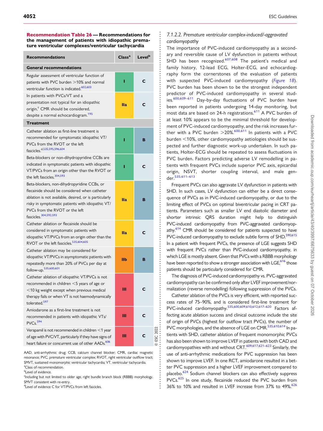
Recommendation Table 26 — Recommendations for the management of patients with idiopathic premature ventricular complexes/ventricular tachycardia

Recommendations Classa Levelb

General recommendations Regular assessment of ventricular function of patients with PVC burden .10% and normal ventricular function is indicated.602,603 I C In patients with PVCs/VT and a presentation not typical for an idiopathic origin,c CMR should be considered, despite a normal echocardiogram.195 IIa C Treatment Catheter ablation as first-line treatment is recommended for symptomatic idiopathic VT/ PVCs from the RVOT or the left fascicles.d,535,595,596,604 I B Beta-blockers or non-dihydropyridine CCBs are indicated in symptomatic patients with idiopathic VT/PVCs from an origin other than the RVOT or the left fascicles.304,593 I C Beta-blockers, non-dihydropyridine CCBs, or flecainide should be considered when catheter ablation is not available, desired, or is particularly risky in symptomatic patients with idiopathic VT/ PVCs from the RVOT or the left fascicles.304,592,593 IIa B Catheter ablation or flecainide should be considered in symptomatic patients with idiopathic VT/PVCs from an origin other than the RVOT or the left fascicles.535,604,605 IIa C Catheter ablation may be considered for idiopathic VT/PVCs in asymptomatic patients with repeatedly more than 20% of PVCs per day at follow-up.535,600,601 IIb B Catheter ablation of idiopathic VT/PVCs is not recommended in children ,5 years of age or ,10 kg weight except when previous medical therapy fails or when VT is not haemodynamically tolerated.597 III C Amiodarone as a first-line treatment is not recommended in patients with idiopathic VTs/ PVCs.594 III C Verapamil is not recommended in children ,1 year of age with PVC/VT, particularly if they have signs of heart failure or concurrent use of other AADs.606 III C © ESC 2022

AAD, anti-arrhythmic drug; CCB, calcium channel blocker; CMR, cardiac magnetic resonance; PVC, premature ventricular complex; RVOT, right ventricular outflow tract; SMVT, sustained monomorphic ventricular tachycardia; VT, ventricular tachycardia. aClass of recommendation. bLevel of evidence. cIncluding but not limited to older age, right bundle branch block (RBBB) morphology, SMVT consistent with re-entry. dLevel of evidence C for VT/PVCs from left fascicles. Downloaded from academic.oup.com/eurheartj/article/43/40/3997/6675633 by guest on 07 October 2026