p.26 抽出テキスト
Any ECG tracing from emergency services, as well as recordings from interrogation of cardiovascular implantable electronic devices (CIEDs) can also contribute to diagnosis.216–219 The resting 12-lead ECG (including high precordial lead)220 is fundamental and should be repeated regularly during recovery. Continuous heart rhythm monitoring is recommended until definite treatment.221,222 Echocardiography may allow early diagnosis to identify any structural abnormality.222,223 Coronary imaging will be important to exclude CAD, dissection, or anomalies.62,224 Coronary optical coherence tomography and/or intravascular ultrasound may be helpful to characterize stenosis/plaque stability and underlying mechanism of stenosis.225 CMR has repeatedly been shown to provide significant incremental diagnostic value, in particular for concealed cardiomyopathy.131,226–228 Primary electrical diseases may be uncovered by provocative manoeuvres such as sodium channel blocker challenge,136,229–231 lying to standing ECGs,232,233 adenosine challenge,144,234 epinephrine challenge,141,152,235–239 ergonovine/ acetylcholine,222,240 mental stress,241,242 and exercise testing.116,117,119,232,243 Electrophysiological study and electroanatomic mapping may be useful to provide patient-specific insights into the mechanism of CA and to offer therapeutic options in some patients.244–248 Genetic testing may identify a molecular cause of SCA by identifying pathogenic mutations in genes associated with specific phenotypes.213,249,250

Recommendation Table 5 — Recommendations for evaluation of sudden cardiac arrest survivors

Recommendations Classa Levelb

Diagnostic evaluation The investigation of a SCA survivor without obvious extra-cardiac cause is recommended to be overseen by a multidisciplinary team.177,251–256 I B In electrically unstable patients after SCA, with suspicion of ongoing myocardial ischaemia, a coronary angiogram is indicated. I C

Continued

ECG during BBR-VT ECG during sinus rhythm

I

II

III

aVR

aVL

aVF

V1

V4

V5

V6

V2

V3 I

II

III

aVR

aVL

aVF

V1

V4

V5

V6

V2

V3

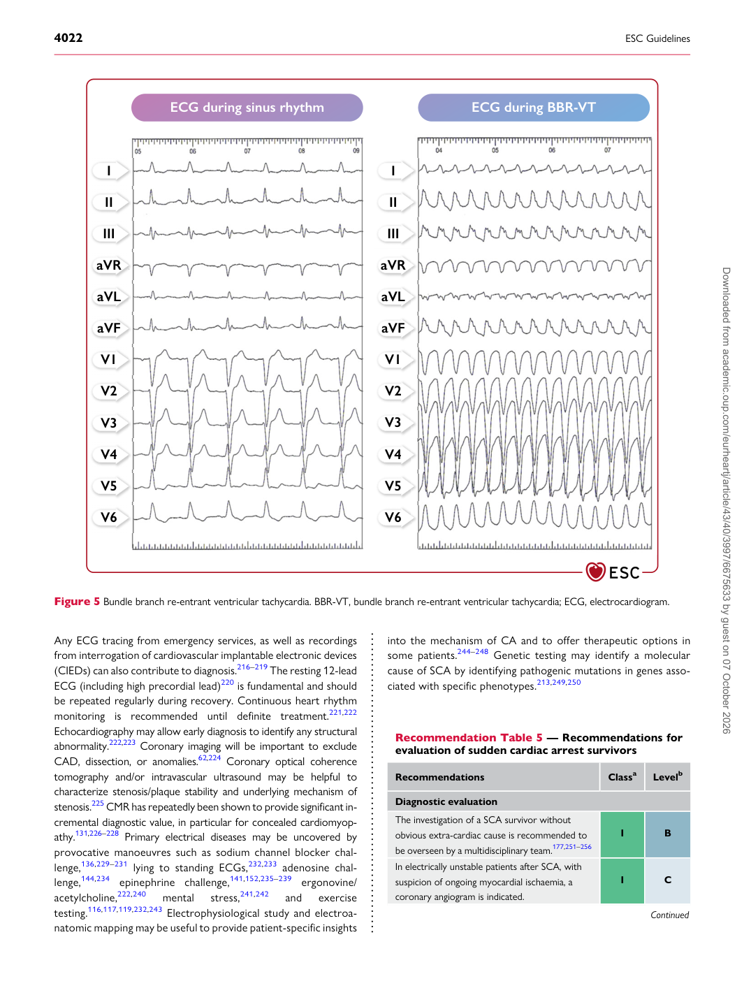
Figure 5 Bundle branch re-entrant ventricular tachycardia. BBR-VT, bundle branch re-entrant ventricular tachycardia; ECG, electrocardiogram. Downloaded from academic.oup.com/eurheartj/article/43/40/3997/6675633 by guest on 07 October 2026