p.25 抽出テキスト
Exclude C AD d

CMR (Class IIa)

Autoim m une se r olo g y

Ca r diac/tissue biopsy

PE T -CT

T

T

M a pping-guided biopsy (if r egional v entricular

i n v ol v ement) (Class IIb)

Go to section Inflammato r y ca r diac diseases

r r

Fu r ther e valuation acco r ding to most li k e

k k l y

diagnosis

Go to section

A R VC

EP study and R V

R R

elect r o-anatomical m a pping (± m a pping-guided biopsy)

(Class IIb) Suggesti v e of SHD other than C AD c

Suggesti v e of C AD b

Suggesti v e of idiopathic origin a

Suggesti v e of inflammato r y disease g

Suggesti v e of other ca r dio m y opat h y f

Suggesti v e of A R VC

R

R e

Go to section Idiopathic PVC/VT C A G

C AD

positi v e

negati v e

No C AD

Go to section C AD

C

C

N Y Y

Patient with first SMVT episode

12-lead ECG

(Class I) Echoca r diogram

(Class I)

VT 12-lead ECG (Cycle length , morpholo

, g y ,

y y

e x e r cise induced)

(Class I) P er sona l h i s t o r y

(C a r d i o vasc ul a r ri sk fac t o r s , sy m p t o m s , co

, -m or bi d iti es , me d i ca ti

a a on ) Fami l y histo r y

(SC D , prima

, r y electrical diseas e , ca

, r dio m y opat h y) Laborato r y

(Elect r o l ytes, (N T -)p

T T r oBN P ,

P P TSH/T4)

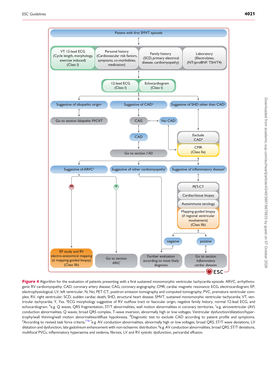
Figure 4 Algorithm for the evaluation of patients presenting with a first sustained monomorphic ventricular tachycardia episode. ARVC, arrhythmogenic RV cardiomyopathy; CAD, coronary artery disease; CAG, coronary angiography; CMR, cardiac magnetic resonance; ECG, electrocardiogram; EP, electrophysiological; LV, left ventricular; N, No; PET-CT, positron emission tomography and computed tomography; PVC, premature ventricular complex; RV, right ventricular; SCD, sudden cardiac death; SHD, structural heart disease; SMVT, sustained monomorphic ventricular tachycardia; VT, ventricular tachycardia; Y, Yes. aECG morphology suggestive of RV outflow tract or fascicular origin, negative family history, normal 12-lead ECG, and echocardiogram. be.g. Q waves, QRS fragmentation, ST/T abnormalities, wall motion abnormalities in coronary territories. ce.g. atrioventricular (AV) conduction abnormalities, Q waves, broad QRS complex, T-wave inversion, abnormally high or low voltages. Ventricular dysfunction/dilatation/hypertrophy/wall thinning/wall motion abnormalities/diffuse hypokinesia. dDiagnostic test to exclude CAD according to patient profile and symptoms. eAccording to revised task force criteria.116 fe.g. AV conduction abnormalities, abnormally high or low voltages, broad QRS, ST/T wave deviations, LV dilatation and dysfunction, late gadolinium enhancement with non-ischaemic distribution. ge.g. AV conduction abnormalities, broad QRS, ST/T deviations, multifocal PVCs, inflammatory hyperaemia and oedema, fibrosis, LV and RV systolic dysfunction, pericardial effusion. Downloaded from academic.oup.com/eurheartj/article/43/40/3997/6675633 by guest on 07 October 2026