p.23 抽出テキスト
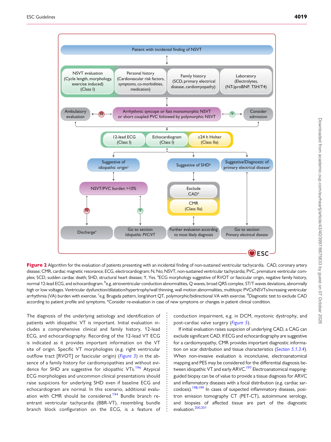
The diagnosis of the underlying aetiology and identification of patients with idiopathic VT is important. Initial evaluation includes a comprehensive clinical and family history, 12-lead ECG, and echocardiography. Recording of the 12-lead VT ECG is indicated as it provides important information on the VT site of origin. Specific VT morphologies (e.g. right ventricular outflow tract [RVOT] or fascicular origin) (Figure 3) in the absence of a family history for cardiomyopathies and without evidence for SHD are suggestive for idiopathic VTs.196 Atypical ECG morphologies and uncommon clinical presentations should raise suspicions for underlying SHD even if baseline ECG and echocardiogram are normal. In this scenario, additional evaluation with CMR should be considered.194 Bundle branch reentrant ventricular tachycardia (BBR-VT), resembling bundle branch block configuration on the ECG, is a feature of conduction impairment, e.g. in DCM, myotonic dystrophy, and post-cardiac valve surgery (Figure 5).

If initial evaluation raises suspicion of underlying CAD, a CAG can exclude significant CAD. If ECG and echocardiography are suggestive for a cardiomyopathy, CMR provides important diagnostic information on scar distribution and tissue characteristics (Section 5.1.3.4). When non-invasive evaluation is inconclusive, electroanatomical mapping and PES may be considered for the differential diagnosis between idiopathic VT and early ARVC.197 Electroanatomical mappingguided biopsy can be of value to provide a tissue diagnosis for ARVC and inflammatory diseases with a focal distribution (e.g. cardiac sarcoidosis).198,199 In cases of suspected inflammatory diseases, positron emission tomography CT (PET-CT), autoimmune serology, and biopsies of affected tissue are part of the diagnostic evaluation.200,201

N

N Patient with incidental finding of NSVT

A r r h ythmic syncope or fast monomorphic NSVT or sho r t coupled PVC f oll

f f o w ed b y po l ymorphic NSVT

Discharge e Go to section

Idiopathic PVC/VT

Suggesti v e of idiopathic origin a

NSVT/PVC bu r den >10%

12-lead ECG

(Class I)

Ambulato r y e valuation Consider

admission

Echoca r diogram

(Class I) ≥ 24 h Holter

(Class IIa)

Suggesti v e of SHD b Suggesti v e/Diagnostic of

prima r y electrical disease c

F u r t he r e va l ua ti

a a on acco r d i n g t o m os t li k e

k k l y d i agnos i s Go to section

P r ima r y elect r ical disease

Y

Y

NSVT e valuation (Cycle length , morpholo

, g y ,

y y

e x e r cise induced)

(Class I) P e r so n a l hi s t o r y

(C a r d i o vascu l a r r

r i sk fac t o r s ,

sym pt oms , co

, - mo r b i d iti es , m e d i ca ti

a a o n ) Fami l y histo r y

(SC D , prima

, r y electrical diseas e , ca

, r dio m y opat h y) Laborato r y

(Elect r o l ytes, (N T -)p

T

T r oBN P ,

P

P TSH/T4)

CMR (Class IIa)

Exclude C AD d

Figure 2 Algorithm for the evaluation of patients presenting with an incidental finding of non-sustained ventricular tachycardia. CAD, coronary artery disease; CMR, cardiac magnetic resonance; ECG, electrocardiogram; N, No; NSVT, non-sustained ventricular tachycardia; PVC, premature ventricular complex; SCD, sudden cardiac death; SHD, structural heart disease; Y, Yes. aECG morphology suggestive of RVOT or fascicular origin, negative family history, normal 12-lead ECG, and echocardiogram. be.g. atrioventricular conduction abnormalities, Q waves, broad QRS complex, ST/T waves deviations, abnormally high or low voltages. Ventricular dysfunction/dilatation/hypertrophy/wall thinning, wall motion abnormalities, multitopic PVCs/NSVTs/increasing ventricular arrhythmia (VA) burden with exercise. ce.g. Brugada pattern, long/short QT, polymorphic/bidirectional VA with exercise. dDiagnostic test to exclude CAD according to patient profile and symptoms. eConsider re-evaluation in case of new symptoms or changes in patient clinical condition. Downloaded from academic.oup.com/eurheartj/article/43/40/3997/6675633 by guest on 07 October 2026