p.18 抽出テキスト
OHCA victims.101–103 Education of public officials and community members regarding the importance of increasing rates of bystander CPR and promoting the use of early defibrillation by lay and professional rescuers is critical to increasing survival rates.

5 Evaluation and treatment. General aspects

5.1 Diagnostic tools

5.1.1. History and physical examination History should focus on ‘red flags’, including features of arrhythmic syncope, e.g. absence of vagal prodrome, and family history of premature or SCD including, e.g. drowning or car accident in long QT syndrome (LQTS), and CPVT.1,106 Subtle features to suggest inherited causes include a family history of epilepsy, sudden infant death syndrome, deafness (LQTS), heart failure, or pacemaker implantation at ,50 years old. Features of diseases related to pro-arrhythmic conditions include mid-systolic click in mitral valve prolapse (MVP) and outflow tract murmurs with Valsalva in HCM. Specific skin features may be relevant, e.g. Lupus pernio, erythema nodosum in sarcoidosis, angiokeratoma in Fabry’s disease, xanthelasma/xanthoma, and palmoplantar keratosis in ARVC.

5.1.2 Laboratory testing

Natriuretic peptides (b-type-natriuretic peptide, or N-terminal pro-b-type-natriuretic peptide) may have a role in the identification of individuals at increased risk of SCD in the general population107,108 or in patients with CAD.109 There is not sufficient evidence to use b-type-natriuretic peptide as a method to select the need for an ICD.110,111

5.1.3 Non-invasive and invasive tests

5.1.3.1. Electrocardiogram and ambulatory electrocardiographic monitoring2 The 12-lead ECG is an important tool for the diagnosis of underlying disease, for risk stratification in selected populations, and for the diagnosis of the VA subtype, if captured. Documentation of arrhythmias related to symptoms is clinically pivotal but may be challenging with sporadic events. The type of ECG-monitoring device and the recording time should therefore match the frequency of clinical events. Monitoring over a period of 24–48 h (typically ‘Holter recording’) is appropriate for daily arrhythmias,112 while intermittent monitoring over a longer period, with patient-activated ECG recorders (or mobile-health/smartphones), should be preferred for infrequent events.113 Implantable loop recorders (ILR) can be useful in diagnosing arrhythmias in patients with potentially life-threatening symptoms, such as unexplained syncope.114

5.1.3.2 Signal-averaged electrocardiogram

Signal-averaged electrocardiogram (SaECG) can detect very low amplitude signals (‘late potentials’) in the terminal QRS segment115 using three time-domain measurements: QRS duration, low-amplitude (,40 μV) signal duration and root mean square voltage of terminal 40 ms QRS.112 Abnormalities in the SaECG can also be assessed by frequency-domain analysis.112 SaECG can contribute to the diagnosis of ARVC.116

5.1.3.3 Exercise testing

Exercise testing is useful for the diagnosis and for evaluating response to therapy in patients with suspected/proven adrenergic-dependent rhythm disturbances, such as exercise-induced idiopathic MVT, PVT, or bidirectional VT in CPVT.117,118 The 4-minute recovery QTc after exercise testing can contribute to the diagnosis LQTS.119

5.1.3.4 Imaging

Imaging is crucial to assess cardiac function and detect cardiomyopathies ( ESC CardioMed chapter 10).120 A negative imaging study supports primary electrical disease in a patient with VA. Echocardiography is a readily available and first-line diagnostic and risk stratification tool for valve diseases, CAD and DCM, HCM, ARVC,121 and left ventricular non-compaction (LVNC). Echocardiographic strainrate imaging allows differentiation between active and passive movement of myocardial segments and early detection of myocardial dysfunction. Wall motion abnormalities can indicate previous infarcts, cardiomyopathies, or inflammatory disease. Global longitudinal strain is a robust measure of left ventricular (LV) function and can detect subtle changes in LV function while LVEF is still preserved.122 Strain imaging can assess mechanical dispersion reflecting inhomogeneous contraction that could be associated with increased risk of VA.122–125 CMR currently provides the most accurate and reproducible measurement of atrial, biventricular global and regional systolic function, and can detect myocardial oedema, fibrosis, infiltration, and

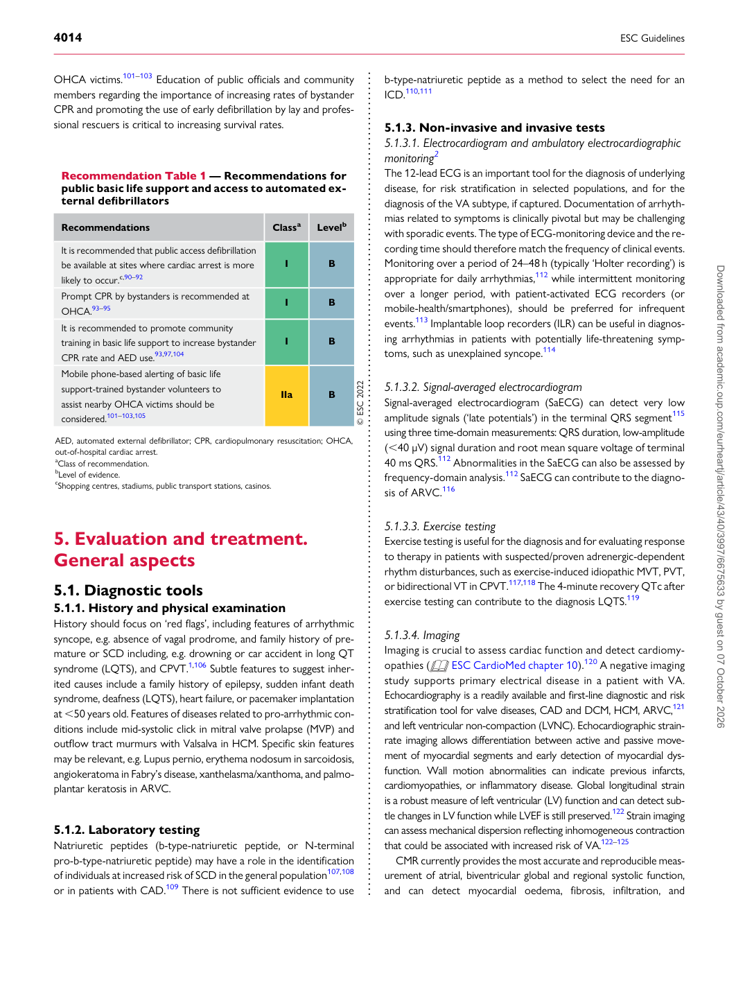
Recommendation Table 1 — Recommendations for public basic life support and access to automated external defibrillators

Recommendations Classa Levelb

It is recommended that public access defibrillation be available at sites where cardiac arrest is more likely to occur.c,90–92 I B Prompt CPR by bystanders is recommended at OHCA.93–95 I B It is recommended to promote community training in basic life support to increase bystander CPR rate and AED use.93,97,104 I B Mobile phone-based alerting of basic life support-trained bystander volunteers to assist nearby OHCA victims should be considered.101–103,105 IIa B © ESC 2022

AED, automated external defibrillator; CPR, cardiopulmonary resuscitation; OHCA, out-of-hospital cardiac arrest. aClass of recommendation. bLevel of evidence. cShopping centres, stadiums, public transport stations, casinos. Downloaded from academic.oup.com/eurheartj/article/43/40/3997/6675633 by guest on 07 October 2026