p.80 抽出テキスト
10.2.4 Multimodal treatment

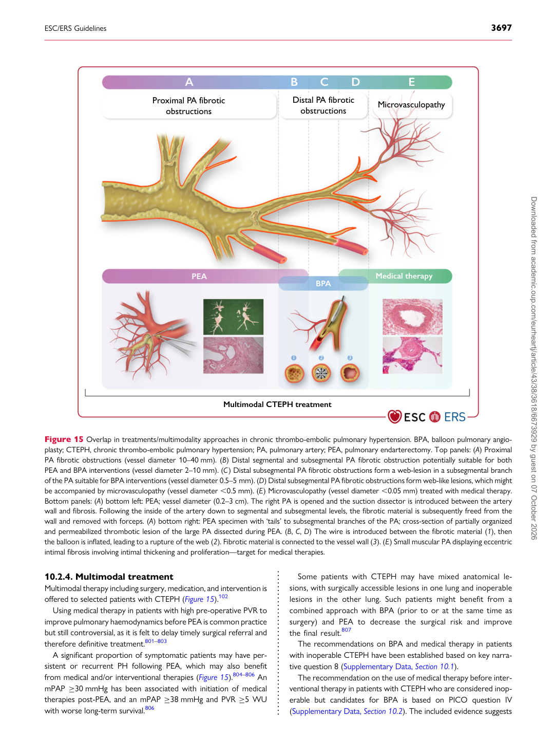
Multimodal therapy including surgery, medication, and intervention is offered to selected patients with CTEPH (Figure 15).102 Using medical therapy in patients with high pre-operative PVR to improve pulmonary haemodynamics before PEA is common practice but still controversial, as it is felt to delay timely surgical referral and therefore definitive treatment.801–803 A significant proportion of symptomatic patients may have persistent or recurrent PH following PEA, which may also benefit from medical and/or interventional therapies (Figure 15).804–806 An mPAP ≥30 mmHg has been associated with initiation of medical therapies post-PEA, and an mPAP ≥38 mmHg and PVR ≥5 WU with worse long-term survival.806 Some patients with CTEPH may have mixed anatomical lesions, with surgically accessible lesions in one lung and inoperable lesions in the other lung. Such patients might benefit from a combined approach with BPA (prior to or at the same time as surgery) and PEA to decrease the surgical risk and improve the final result.807

The recommendations on BPA and medical therapy in patients with inoperable CTEPH have been established based on key narrative question 8 (Supplementary Data, Section 10.1). The recommendation on the use of medical therapy before interventional therapy in patients with CTEPH who are considered inoperable but candidates for BPA is based on PICO question IV (Supplementary Data, Section 10.2). The included evidence suggests

A B C D E

PEA Medical therapy

Microvasculopathy Proximal PA fibrotic

obstructions

Multimodal CTEPH treatment

Distal PA fibrotic

obstructions

BPA

1 2 3

Figure 15 Overlap in treatments/multimodality approaches in chronic thrombo-embolic pulmonary hypertension. BPA, balloon pulmonary angioplasty; CTEPH, chronic thrombo-embolic pulmonary hypertension; PA, pulmonary artery; PEA, pulmonary endarterectomy. Top panels: (A) Proximal PA fibrotic obstructions (vessel diameter 10–40 mm). (B) Distal segmental and subsegmental PA fibrotic obstruction potentially suitable for both PEA and BPA interventions (vessel diameter 2–10 mm). (C) Distal subsegmental PA fibrotic obstructions form a web-lesion in a subsegmental branch of the PA suitable for BPA interventions (vessel diameter 0.5–5 mm). (D) Distal subsegmental PA fibrotic obstructions form web-like lesions, which might be accompanied by microvasculopathy (vessel diameter ,0.5 mm). (E) Microvasculopathy (vessel diameter ,0.05 mm) treated with medical therapy. Bottom panels: (A) bottom left: PEA; vessel diameter (0.2–3 cm). The right PA is opened and the suction dissector is introduced between the artery wall and fibrosis. Following the inside of the artery down to segmental and subsegmental levels, the fibrotic material is subsequently freed from the wall and removed with forceps. (A) bottom right: PEA specimen with ‘tails’ to subsegmental branches of the PA; cross-section of partially organized and permeabilized thrombotic lesion of the large PA dissected during PEA. (B, C, D) The wire is introduced between the fibrotic material (1), then the balloon is inflated, leading to a rupture of the web (2). Fibrotic material is connected to the vessel wall (3). (E) Small muscular PA displaying eccentric intimal fibrosis involving intimal thickening and proliferation—target for medical therapies. Downloaded from academic.oup.com/eurheartj/article/43/38/3618/6673929 by guest on 07 October 2026