p.77 抽出テキスト
considered to have CTEPD with or without PH; CTEPH remains the preferred term for patients with PH, as defined in Section 3.1 (Table 5).54 Chronic thrombo-embolic pulmonary disease describes symptomatic patients with mismatched perfusion defects on V/Q scan and with signs of chronic, organized, fibrotic clots on CTPA or DSA, such as ring-like stenoses, webs/slits, and chronic total occlusions (pouch lesions or tapered lesions), after at least 3 months of therapeutic anticoagulation. Pulmonary hypertension in this setting is not only a consequence of PA obstruction by organized fibrotic clots but can also be related to the associated microvasculopathy. In those patients without PH at rest, breathlessness could be due to exercise PH (see definition in Section 3.1, Table 5) and/or increased dead space ventilation.54 Excluding ventilatory limitation, deconditioning and psychogenic hyperventilation syndrome by CPET and LV myocardial or valvular disease by echocardiography is of upmost importance when making therapeutic decisions in patients with CTEPD without PH.

10.1 Diagnosis

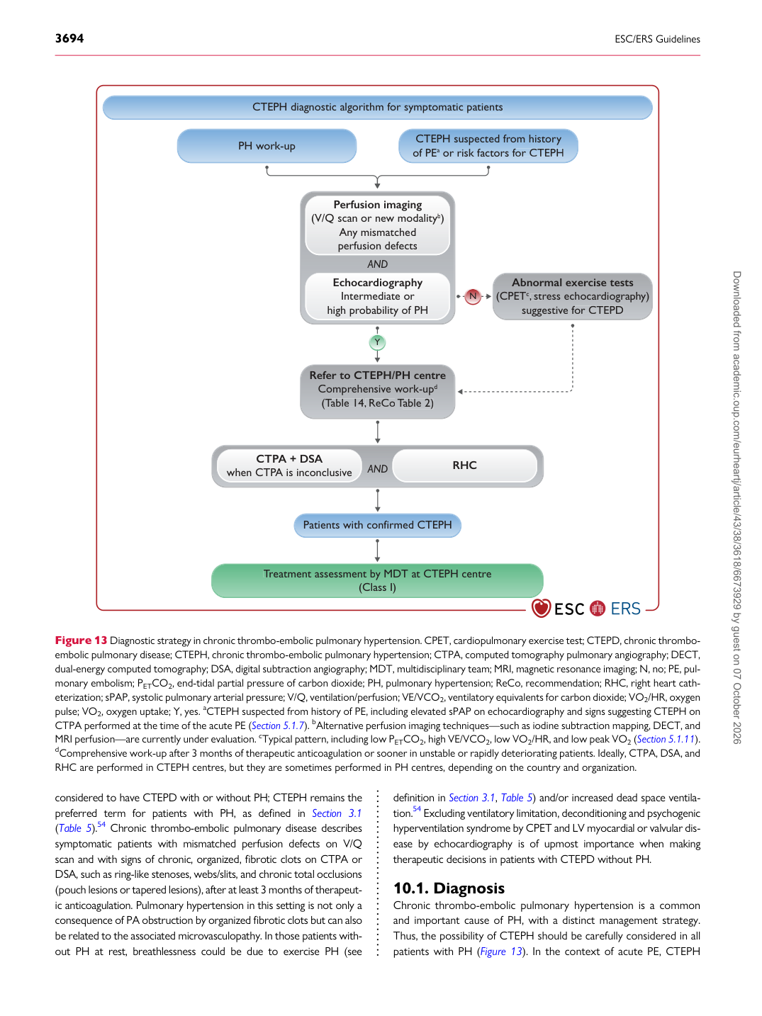
Chronic thrombo-embolic pulmonary hypertension is a common and important cause of PH, with a distinct management strategy. Thus, the possibility of CTEPH should be carefully considered in all patients with PH (Figure 13). In the context of acute PE, CTEPH

CTEPH diagnostic al g orithm f or symptomatic patients

f f

Patients with confirmed CTEPH

CTEPH suspected f r om histo r y of PE a or risk factors f or CTEPH

f f

T r T T eatment assessment b y MDT at CTEPH cent r e

(Class I)

PH w ork-up

N

Y

P erfusion imaging (V/Q scan or n e w modality b )

A n y mismatched perfusion de f ects

f f

Echoca r diography

Intermediate or high p r obability of PH

R efer to CTEPH/PH cent r e

Comp r ehensi v e w ork-up d ( T able 14 T T , ReCo

, T able 2)

T T Abnormal exe r cise tests

(CPET c , st

, r ess echoca r diogr a p h y) suggesti v e f or CTEPD

f f

AN D

CT P A + D

P P S A when CT P A is inconclusi

P P v e RHC

AN D

Figure 13 Diagnostic strategy in chronic thrombo-embolic pulmonary hypertension. CPET, cardiopulmonary exercise test; CTEPD, chronic thromboembolic pulmonary disease; CTEPH, chronic thrombo-embolic pulmonary hypertension; CTPA, computed tomography pulmonary angiography; DECT, dual-energy computed tomography; DSA, digital subtraction angiography; MDT, multidisciplinary team; MRI, magnetic resonance imaging; N, no; PE, pulmonary embolism; PETCO2, end-tidal partial pressure of carbon dioxide; PH, pulmonary hypertension; ReCo, recommendation; RHC, right heart catheterization; sPAP, systolic pulmonary arterial pressure; V/Q, ventilation/perfusion; VE/VCO2, ventilatory equivalents for carbon dioxide; VO2/HR, oxygen pulse; VO2, oxygen uptake; Y, yes. aCTEPH suspected from history of PE, including elevated sPAP on echocardiography and signs suggesting CTEPH on CTPA performed at the time of the acute PE (Section 5.1.7). bAlternative perfusion imaging techniques—such as iodine subtraction mapping, DECT, and MRI perfusion—are currently under evaluation. cTypical pattern, including low PETCO2, high VE/VCO2, low VO2/HR, and low peak VO2 (Section 5.1.11). dComprehensive work-up after 3 months of therapeutic anticoagulation or sooner in unstable or rapidly deteriorating patients. Ideally, CTPA, DSA, and RHC are performed in CTEPH centres, but they are sometimes performed in PH centres, depending on the country and organization. Downloaded from academic.oup.com/eurheartj/article/43/38/3618/6673929 by guest on 07 October 2026