p.74 抽出テキスト
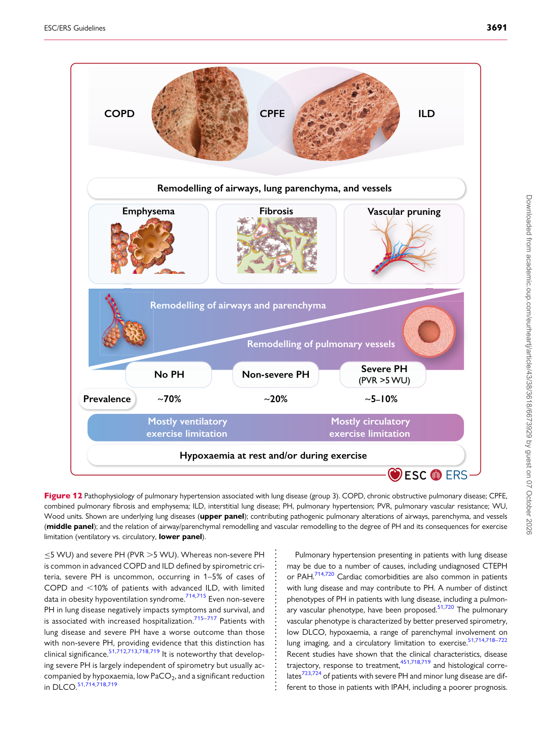
≤5 WU) and severe PH (PVR .5 WU). Whereas non-severe PH is common in advanced COPD and ILD defined by spirometric criteria, severe PH is uncommon, occurring in 1–5% of cases of COPD and ,10% of patients with advanced ILD, with limited data in obesity hypoventilation syndrome.714,715 Even non-severe PH in lung disease negatively impacts symptoms and survival, and is associated with increased hospitalization.715–717 Patients with lung disease and severe PH have a worse outcome than those with non-severe PH, providing evidence that this distinction has clinical significance.51,712,713,718,719 It is noteworthy that developing severe PH is largely independent of spirometry but usually accompanied by hypoxaemia, low PaCO2, and a significant reduction in DLCO.51,714,718,719 Pulmonary hypertension presenting in patients with lung disease may be due to a number of causes, including undiagnosed CTEPH or PAH.714,720 Cardiac comorbidities are also common in patients with lung disease and may contribute to PH. A number of distinct phenotypes of PH in patients with lung disease, including a pulmonary vascular phenotype, have been proposed.51,720 The pulmonary vascular phenotype is characterized by better preserved spirometry, low DLCO, hypoxaemia, a range of parenchymal involvement on lung imaging, and a circulatory limitation to exercise.51,714,718–722

Recent studies have shown that the clinical characteristics, disease trajectory, response to treatment,451,718,719 and histological correlates723,724 of patients with severe PH and minor lung disease are different to those in patients with IPAH, including a poorer prognosis.

Remodelling of airways, lung parenchyma, and vessels

Prevalence ~70% ~20% ~5–10%

Mostly ventilatory exercise limitation Mostly circulatory exercise limitation

Remodelling of pulmonary vessels

Remodelling of airways and parenchyma

Hypoxaemia at rest and/or during exercise

No PH Non-severe PH Severe PH (PVR >5 WU)

COPD ILD

CPFE

R

Emphysema Fibrosis Vascular pruning

C

CPFE

Figure 12 Pathophysiology of pulmonary hypertension associated with lung disease (group 3). COPD, chronic obstructive pulmonary disease; CPFE, combined pulmonary fibrosis and emphysema; ILD, interstitial lung disease; PH, pulmonary hypertension; PVR, pulmonary vascular resistance; WU, Wood units. Shown are underlying lung diseases (upper panel); contributing pathogenic pulmonary alterations of airways, parenchyma, and vessels (middle panel); and the relation of airway/parenchymal remodelling and vascular remodelling to the degree of PH and its consequences for exercise limitation (ventilatory vs. circulatory, lower panel). Downloaded from academic.oup.com/eurheartj/article/43/38/3618/6673929 by guest on 07 October 2026