p.71 抽出テキスト
with a rapid increase in PAWP when challenged with increased systemic venous return.674 While the upper limit of normal remains controversial,142,143,665,667 a PAWP cut-off of .18 mmHg has been suggested to identify HFpEF as the underlying cause of PH, despite normal PAWP at baseline.143 While this may help to classify PH, therapeutic consequences of such testing remain to be determined. As differentiating between severe PH associated with HFpEF and IPAH with cardiac comorbidities is challenging, patients with an unclear diagnosis, particularly those with a predominant pre-capillary component (e.g. PVR .5 WU), should be referred to a PH centre for individualized management.

8.3 Therapy

The primary strategy in managing PH-LHD is optimizing treatment of the underlying cardiac disease. Nevertheless, a pathophysiological sequence ranging from left-sided heart disease via pulmonary circulation to chronic right heart strain (at rest or exercise) is present in many patients.47 Since deterioration of RV function over time is associated with poor outcomes in HFpEF,658 preserving RV function should be considered an important treatment goal. Diuretics remain the cornerstone of medical therapy in the presence of fluid retention due to PH-LHD. There is limited and conflicting evidence for the use of drugs approved for PAH in patients with group 2 PH. Some medications may have variable and potentially detrimental effects in such patients and are therefore not indicated in PH-LHD. Management strategies for PH in various left heart aetiologies are described below.

8.3.1 Pulmonary hypertension associated with

left-sided heart failure 8.3.1.1. Heart failure with reduced ejection fraction Patients with HFrEF or HFmrEF require guideline-directed treatment including established medical and interventional therapies.27 In patients with advanced HFrEF, implanting an LVAD may significantly reduce or even normalize mPAP,675 although this is not achieved in all patients,676 and an increased DPG emerged as a negative prognostic factor after LVAD implantation.677 With regards to PAH drugs, bosentan was assessed in an RCT of patients with PH associated with HFrEF,678 showing no efficacy but an increase in adverse events compared with placebo, predominantly related to fluid retention. Small studies have suggested that sildenafil may improve haemodynamics and exercise capacity in PH and HFrEF,679–681 but RCTs are lacking.

8.3.1.2 Heart failure with preserved ejection fraction

In patients with HFpEF, blood pressure, volume load, and risk factors should be controlled, which may lower filling pressures and PAP.27 Recently, the SGLT-2i empagliflozin improved outcomes in patients

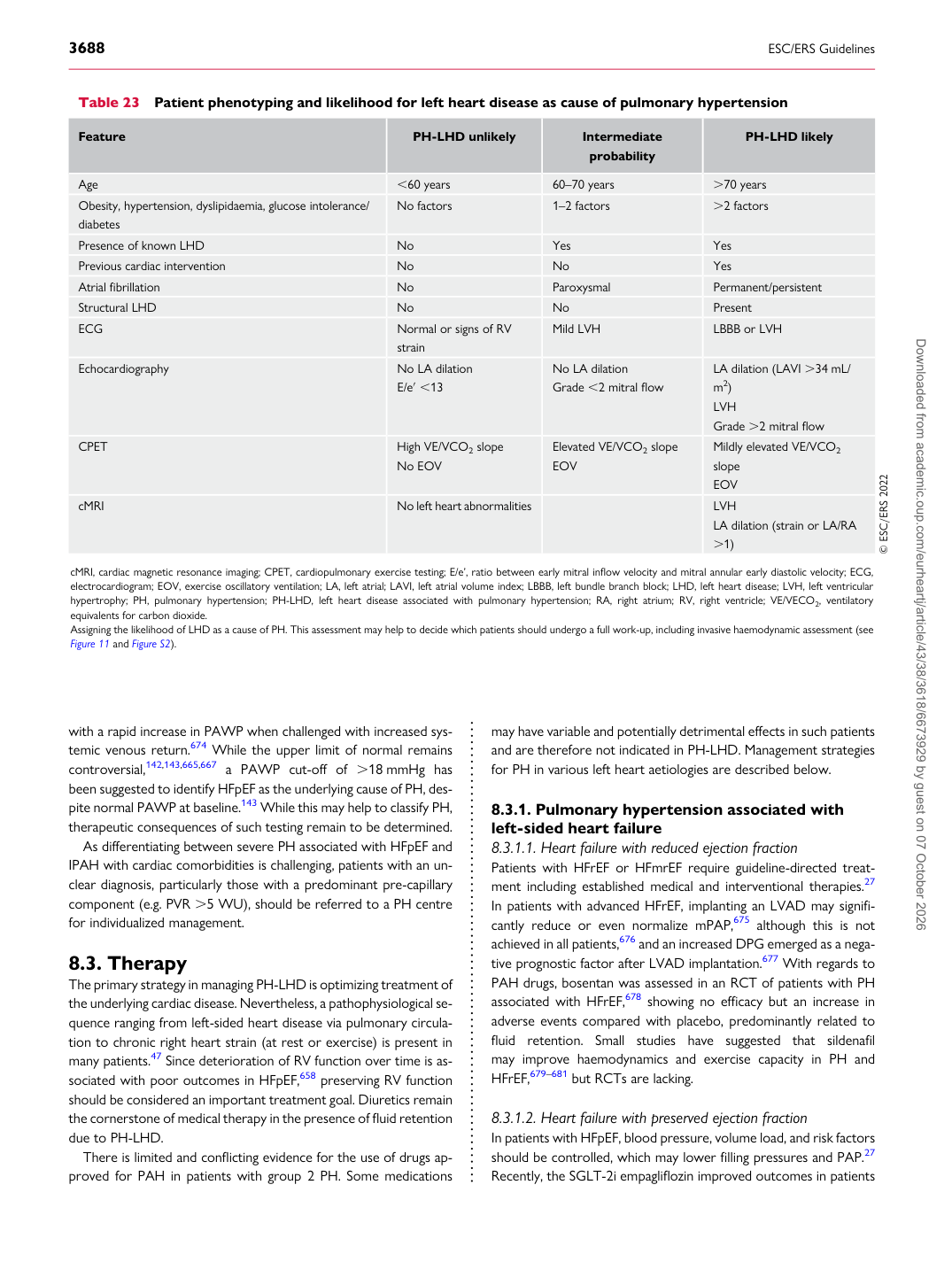
Table 23 Patient phenotyping and likelihood for left heart disease as cause of pulmonary hypertension

Feature PH-LHD unlikely Intermediate

probability PH-LHD likely

Age ,60 years 60–70 years .70 years Obesity, hypertension, dyslipidaemia, glucose intolerance/ diabetes No factors 1–2 factors .2 factors Presence of known LHD No Yes Yes Previous cardiac intervention No No Yes Atrial fibrillation No Paroxysmal Permanent/persistent Structural LHD No No Present ECG Normal or signs of RV

strain Mild LVH LBBB or LVH

Echocardiography No LA dilation

E/e′ ,13 No LA dilation

Grade ,2 mitral flow LA dilation (LAVI .34 mL/

m2) LVH Grade .2 mitral flow

CPET High VE/VCO2 slope

No EOV Elevated VE/VCO2 slope

EOV Mildly elevated VE/VCO2 slope EOV

cMRI No left heart abnormalities LVH LA dilation (strain or LA/RA

.1) © ESC/ERS 2022

cMRI, cardiac magnetic resonance imaging; CPET, cardiopulmonary exercise testing; E/e’, ratio between early mitral inflow velocity and mitral annular early diastolic velocity; ECG, electrocardiogram; EOV, exercise oscillatory ventilation; LA, left atrial; LAVI, left atrial volume index; LBBB, left bundle branch block; LHD, left heart disease; LVH, left ventricular hypertrophy; PH, pulmonary hypertension; PH-LHD, left heart disease associated with pulmonary hypertension; RA, right atrium; RV, right ventricle; VE/VECO2, ventilatory equivalents for carbon dioxide. Assigning the likelihood of LHD as a cause of PH. This assessment may help to decide which patients should undergo a full work-up, including invasive haemodynamic assessment (see Figure 11 and Figure S2). Downloaded from academic.oup.com/eurheartj/article/43/38/3618/6673929 by guest on 07 October 2026