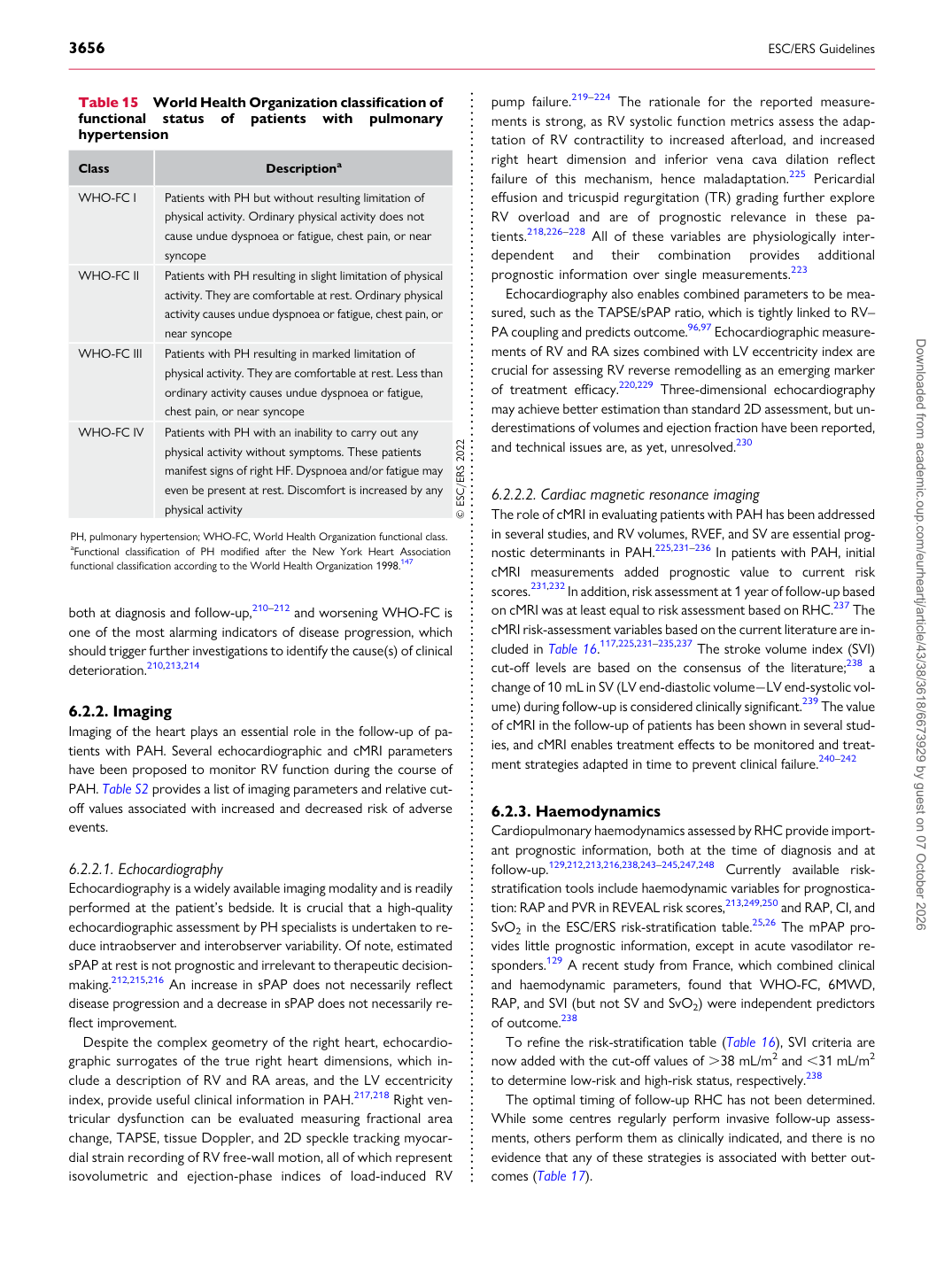
p.39 抽出テキスト
both at diagnosis and follow-up,210–212 and worsening WHO-FC is one of the most alarming indicators of disease progression, which should trigger further investigations to identify the cause(s) of clinical deterioration.210,213,214

6.2.2 Imaging

Imaging of the heart plays an essential role in the follow-up of patients with PAH. Several echocardiographic and cMRI parameters have been proposed to monitor RV function during the course of PAH. Table S2 provides a list of imaging parameters and relative cutoff values associated with increased and decreased risk of adverse events.

6.2.2.1 Echocardiography

Echocardiography is a widely available imaging modality and is readily performed at the patient’s bedside. It is crucial that a high-quality echocardiographic assessment by PH specialists is undertaken to reduce intraobserver and interobserver variability. Of note, estimated sPAP at rest is not prognostic and irrelevant to therapeutic decisionmaking.212,215,216 An increase in sPAP does not necessarily reflect disease progression and a decrease in sPAP does not necessarily reflect improvement. Despite the complex geometry of the right heart, echocardiographic surrogates of the true right heart dimensions, which include a description of RV and RA areas, and the LV eccentricity index, provide useful clinical information in PAH.217,218 Right ventricular dysfunction can be evaluated measuring fractional area change, TAPSE, tissue Doppler, and 2D speckle tracking myocardial strain recording of RV free-wall motion, all of which represent isovolumetric and ejection-phase indices of load-induced RV pump failure.219–224 The rationale for the reported measurements is strong, as RV systolic function metrics assess the adaptation of RV contractility to increased afterload, and increased right heart dimension and inferior vena cava dilation reflect failure of this mechanism, hence maladaptation.225 Pericardial effusion and tricuspid regurgitation (TR) grading further explore RV overload and are of prognostic relevance in these patients.218,226–228 All of these variables are physiologically interdependent and their combination provides additional prognostic information over single measurements.223

Echocardiography also enables combined parameters to be measured, such as the TAPSE/sPAP ratio, which is tightly linked to RV– PA coupling and predicts outcome.96,97 Echocardiographic measurements of RV and RA sizes combined with LV eccentricity index are crucial for assessing RV reverse remodelling as an emerging marker of treatment efficacy.220,229 Three-dimensional echocardiography may achieve better estimation than standard 2D assessment, but underestimations of volumes and ejection fraction have been reported, and technical issues are, as yet, unresolved.230

6.2.2.2 Cardiac magnetic resonance imaging

The role of cMRI in evaluating patients with PAH has been addressed in several studies, and RV volumes, RVEF, and SV are essential prognostic determinants in PAH.225,231–236 In patients with PAH, initial cMRI measurements added prognostic value to current risk scores.231,232 In addition, risk assessment at 1 year of follow-up based on cMRI was at least equal to risk assessment based on RHC.237 The cMRI risk-assessment variables based on the current literature are included in Table 16.117,225,231–235,237 The stroke volume index (SVI) cut-off levels are based on the consensus of the literature;238 a change of 10 mL in SV (LV end-diastolic volume−LV end-systolic volume) during follow-up is considered clinically significant.239 The value of cMRI in the follow-up of patients has been shown in several studies, and cMRI enables treatment effects to be monitored and treatment strategies adapted in time to prevent clinical failure.240–242

6.2.3 Haemodynamics

Cardiopulmonary haemodynamics assessed by RHC provide important prognostic information, both at the time of diagnosis and at follow-up.129,212,213,216,238,243–245,247,248 Currently available riskstratification tools include haemodynamic variables for prognostication: RAP and PVR in REVEAL risk scores,213,249,250 and RAP, CI, and SvO2 in the ESC/ERS risk-stratification table.25,26 The mPAP provides little prognostic information, except in acute vasodilator responders.129 A recent study from France, which combined clinical and haemodynamic parameters, found that WHO-FC, 6MWD, RAP, and SVI (but not SV and SvO2) were independent predictors of outcome.238 To refine the risk-stratification table (Table 16), SVI criteria are now added with the cut-off values of .38 mL/m2 and ,31 mL/m2 to determine low-risk and high-risk status, respectively.238 The optimal timing of follow-up RHC has not been determined. While some centres regularly perform invasive follow-up assessments, others perform them as clinically indicated, and there is no evidence that any of these strategies is associated with better outcomes (Table 17).

Table 15 World Health Organization classification of functional status of patients with pulmonary hypertension

Class Descriptiona

WHO-FC I Patients with PH but without resulting limitation of physical activity. Ordinary physical activity does not

cause undue dyspnoea or fatigue, chest pain, or near syncope

WHO-FC II Patients with PH resulting in slight limitation of physical activity. They are comfortable at rest. Ordinary physical

activity causes undue dyspnoea or fatigue, chest pain, or near syncope

WHO-FC III Patients with PH resulting in marked limitation of

physical activity. They are comfortable at rest. Less than ordinary activity causes undue dyspnoea or fatigue, chest pain, or near syncope

WHO-FC IV Patients with PH with an inability to carry out any

physical activity without symptoms. These patients manifest signs of right HF. Dyspnoea and/or fatigue may even be present at rest. Discomfort is increased by any physical activity © ESC/ERS 2022

PH, pulmonary hypertension; WHO-FC, World Health Organization functional class. aFunctional classification of PH modified after the New York Heart Association functional classification according to the World Health Organization 1998.147 Downloaded from academic.oup.com/eurheartj/article/43/38/3618/6673929 by guest on 07 October 2026