p.37 抽出テキスト
systematic review and meta-analysis reported a CTEPH incidence of 0.6% in all patients with acute PE, 3.2% in survivors, and 2.8% in survivors without major comorbidities.178 A multicentre, observational, screening study reported a CTEPH incidence of 3.7/1000 patient-years and a 2 year cumulative incidence of 0.79% following acute PE.200 A recent prospective observational study (FOCUS, Follow-up After Acute Pulmonary Embolism) showed a cumulative 2 year incidence of 2.3% and 16.0% for CTEPH and post-PE impairment, respectively, which were both associated with a higher risk of rehospitalization and death.201 Due to insufficient awareness, some patients may have a delayed diagnosis of CTEPH because they may initially be misclassified as acute PE.112 In this context, the current guidelines do not recommend routine follow-up of patients with PE by imaging methods of the pulmonary vascular tree, but suggest evaluating the index imaging test used to diagnose acute PE for signs of CTEPH. Echocardiography is the preferred first-line diagnostic test in patients with suspected CTEPH.103 Up to 50% of patients have persistent perfusion defects after an acute PE; however, the clinical relevance is unclear.202–204 All patients in whom symptoms can be attributed to post-thrombotic deposits within PAs are considered to have CTEPD, with or without PH.54 While persistent dyspnoea is common after acute PE,205 the prevalence of CTEPD without PH is unknown and requires further study (see Section 10.1). A study exploring screening for CTEPH following acute PE identified, using echocardiography, a low yield of additional CTEPH diagnoses in asymptomatic patients.206 Current PE guidelines recommend that further diagnostic evaluation may be considered in asymptomatic patients with risk factors for CTEPH at 3–6 months’ follow-up.103,207 Approaches to early detection of CTEPH following acute PE are based on identifying patients at increased risk.208 In patients with persistent or new-onset dyspnoea after PE, non-invasive approaches use echocardiography to assess for PH and cross-sectional imaging to assess for persistent perfusion defects. Limited data exist on strategies using DECT, CT lung subtraction iodine mapping, or 3D MR perfusion imaging. Scoring systems, including the Leiden CTEPH rule-out criteria206,209 can be used to inform diagnostic strategies. Cardiopulmonary exercise testing may identify characteristic features of exercise limitation due to PVD, or suggest an alternative diagnosis. The optimal timing for assessing symptoms to aid early detection of CTEPH may be 3–6 months after acute PE, coinciding with the routine evaluation of anticoagulant treatment, but earlier assessment may be necessary in highly symptomatic or deteriorating patients.54,103

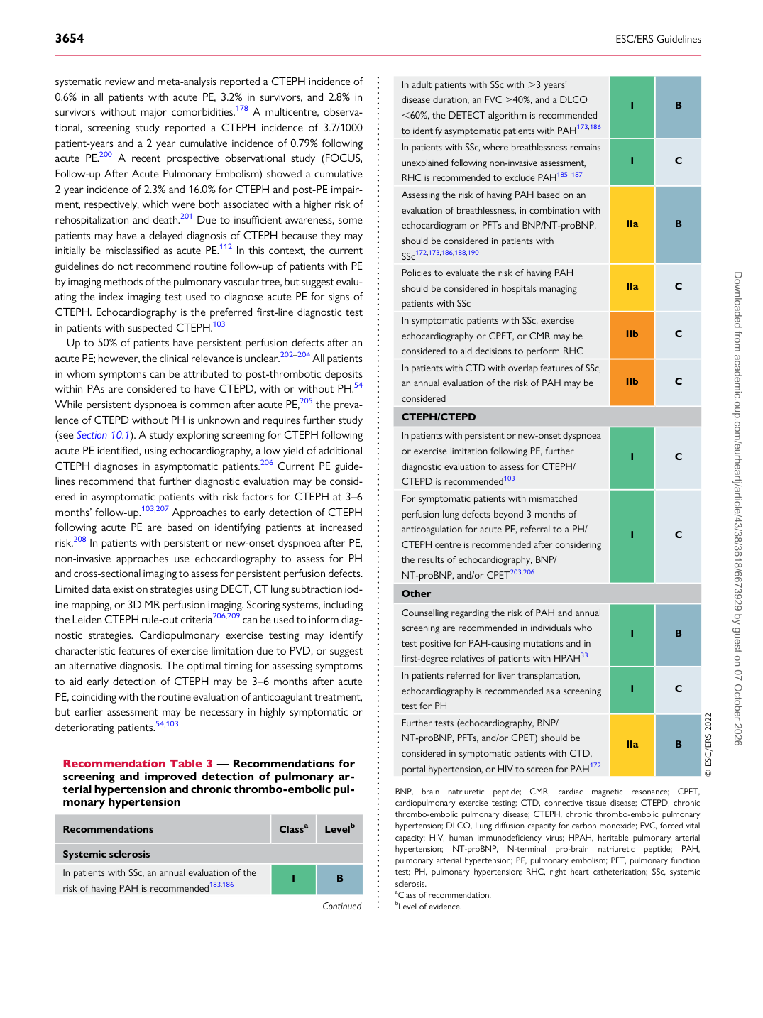
Recommendation Table 3 — Recommendations for screening and improved detection of pulmonary arterial hypertension and chronic thrombo-embolic pulmonary hypertension

Recommendations Classa Levelb

Systemic sclerosis In patients with SSc, an annual evaluation of the risk of having PAH is recommended183,186 I B

Continued In adult patients with SSc with .3 years’

disease duration, an FVC ≥40%, and a DLCO ,60%, the DETECT algorithm is recommended to identify asymptomatic patients with PAH173,186 I B In patients with SSc, where breathlessness remains unexplained following non-invasive assessment, RHC is recommended to exclude PAH185–187 I C Assessing the risk of having PAH based on an evaluation of breathlessness, in combination with echocardiogram or PFTs and BNP/NT-proBNP, should be considered in patients with SSc172,173,186,188,190 IIa B Policies to evaluate the risk of having PAH should be considered in hospitals managing patients with SSc IIa C In symptomatic patients with SSc, exercise echocardiography or CPET, or CMR may be considered to aid decisions to perform RHC IIb C In patients with CTD with overlap features of SSc, an annual evaluation of the risk of PAH may be considered IIb C CTEPH/CTEPD In patients with persistent or new-onset dyspnoea or exercise limitation following PE, further diagnostic evaluation to assess for CTEPH/ CTEPD is recommended103 I C For symptomatic patients with mismatched perfusion lung defects beyond 3 months of anticoagulation for acute PE, referral to a PH/ CTEPH centre is recommended after considering the results of echocardiography, BNP/ NT-proBNP, and/or CPET203,206 I C Other Counselling regarding the risk of PAH and annual screening are recommended in individuals who test positive for PAH-causing mutations and in first-degree relatives of patients with HPAH33 I B In patients referred for liver transplantation, echocardiography is recommended as a screening test for PH I C Further tests (echocardiography, BNP/ NT-proBNP, PFTs, and/or CPET) should be considered in symptomatic patients with CTD, portal hypertension, or HIV to screen for PAH172 IIa B © ESC/ERS 2022 BNP, brain natriuretic peptide; CMR, cardiac magnetic resonance; CPET, cardiopulmonary exercise testing; CTD, connective tissue disease; CTEPD, chronic thrombo-embolic pulmonary disease; CTEPH, chronic thrombo-embolic pulmonary hypertension; DLCO, Lung diffusion capacity for carbon monoxide; FVC, forced vital capacity; HIV, human immunodeficiency virus; HPAH, heritable pulmonary arterial hypertension; NT-proBNP, N-terminal pro-brain natriuretic peptide; PAH, pulmonary arterial hypertension; PE, pulmonary embolism; PFT, pulmonary function test; PH, pulmonary hypertension; RHC, right heart catheterization; SSc, systemic sclerosis. aClass of recommendation. bLevel of evidence. Downloaded from academic.oup.com/eurheartj/article/43/38/3618/6673929 by guest on 07 October 2026