p.35 抽出テキスト
(WHO-FC III/IV), clinical signs of RV failure, syncope, signs of low CO state, poorly tolerated arrhythmias, and compromised or deteriorated haemodynamic status (hypotension, tachycardia). Such cases must be immediately managed as inpatients for initial work-up at a nearby hospital or PH centre. The presence of RV dysfunction by echocardiography, elevated levels of cardiac biomarkers, and/or haemodynamic instability must prompt referral to a PH centre for immediate assessment. This diagnostic process emphasizes the importance of sufficient awareness and collaboration between first-line, specialized medicine and PH centres. Effective and rapid collaboration between each partner permits earlier diagnosis and management, and improves outcomes.

5.3 Screening and early detection

Despite the advent of PAH therapies that prevent clinical worsening166–168 and effective interventions for CTEPH,102 the time from symptom onset to PH diagnosis remains at .2 years,169,170 with most patients presenting with advanced disease. Decreasing the time to diagnosis may reduce emotional uncertainty in patients,171 reduce the use of health care resources, and enable treatment at an earlier stage when therapies may be more effective.172 A proposed multifaceted approach172 to facilitate an earlier diagnosis includes: (1) screening asymptomatic, high-risk groups (with

exercise EOV increasing during

exercise exercise

No EOV

5.1.12 Right heart

catheterization Pre-capillary PH Post-capillary PH Pre-capillary PH Pre- (or post-) capillary

PH © ESC/ERS 2022

ABG, arterial blood gas analysis; CT, computed tomography; CTEPH, chronic thrombo-embolic pulmonary hypertension; DLCO, Lung diffusion capacity for carbon monoxide; EOV, exercise oscillatory ventilation; HFpEF, heart failure with preserved ejection fraction; HFrEF, heart failure with reduced ejection fraction; IPAH, idiopathic pulmonary arterial hypertension; LA, left atrium; LHD, left heart disease; LV, left ventricle; PA, pulmonary artery; PaCO2, partial pressure of arterial carbon dioxide; PAH, pulmonary arterial hypertension; PaO2, partial pressure of arterial oxygen; PETCO2, end-tidal partial pressure of carbon dioxide; PFT, pulmonary function test; PH, pulmonary hypertension; PVOD, pulmonary veno-occlusive disease; RA, right atrium; RV, right ventricle; sPAP, systolic pulmonary arterial pressure; SPECT, single-photon emission computed tomography; SSc-PAH, systemic sclerosis-associated pulmonary arterial hypertension; VE/VCO2, ventilatory equivalent for carbon dioxide; V/Q, ventilation perfusion scintigraphy; VTE, venous thrombo-embolism. ↓, reduced; ↑, increased. aHowever, it may affect individuals of all ages and sexes; diagnosis in males should not be delayed.

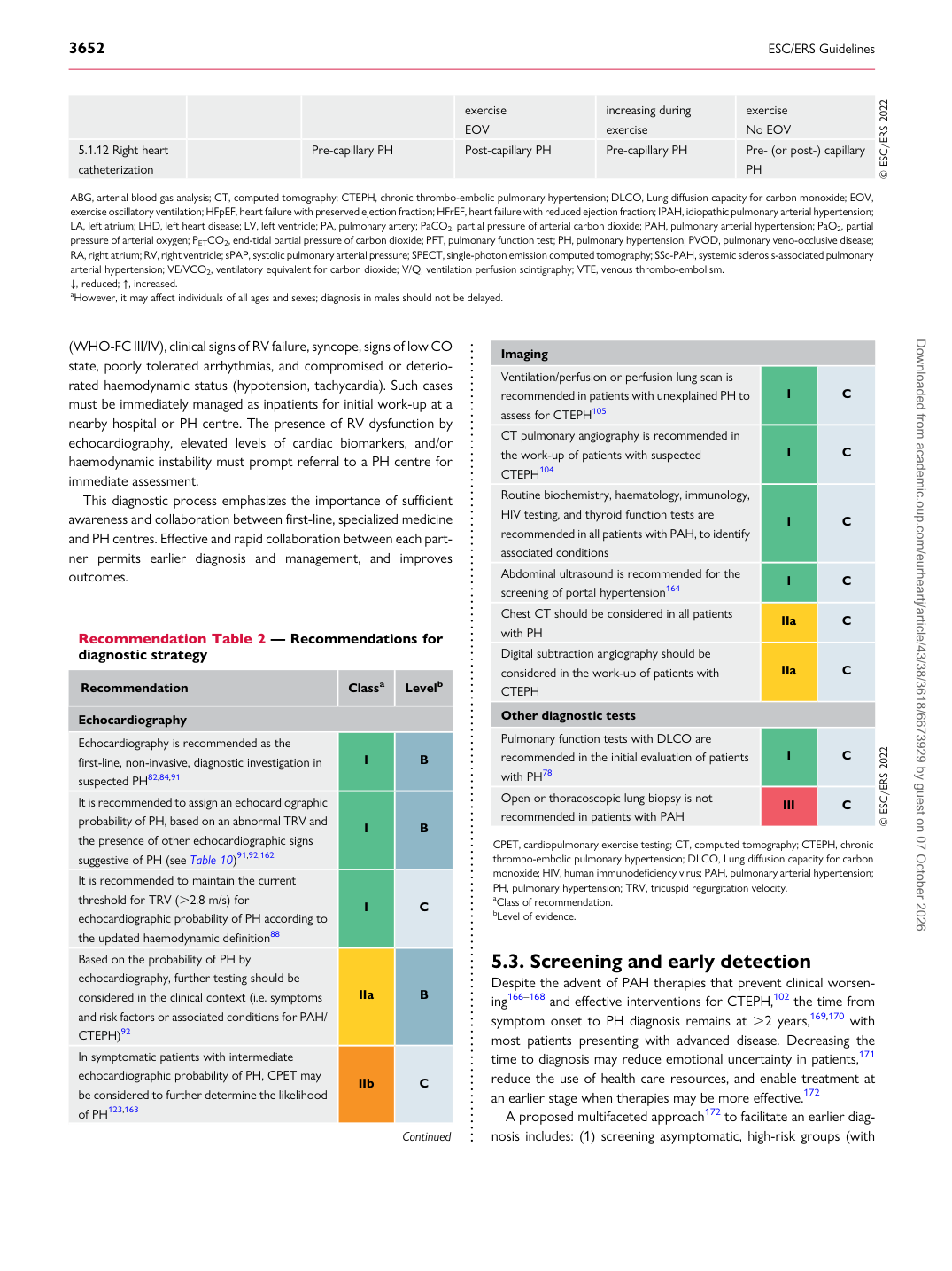
Recommendation Table 2 — Recommendations for diagnostic strategy

Recommendation Classa Levelb

Echocardiography Echocardiography is recommended as the first-line, non-invasive, diagnostic investigation in suspected PH82,84,91 I B It is recommended to assign an echocardiographic probability of PH, based on an abnormal TRV and the presence of other echocardiographic signs suggestive of PH (see Table 10)91,92,162 I B It is recommended to maintain the current threshold for TRV (.2.8 m/s) for echocardiographic probability of PH according to the updated haemodynamic definition88 I C Based on the probability of PH by echocardiography, further testing should be considered in the clinical context (i.e. symptoms and risk factors or associated conditions for PAH/ CTEPH)92 IIa B In symptomatic patients with intermediate echocardiographic probability of PH, CPET may be considered to further determine the likelihood of PH123,163 IIb C

Continued Imaging

Ventilation/perfusion or perfusion lung scan is recommended in patients with unexplained PH to assess for CTEPH105 I C CT pulmonary angiography is recommended in the work-up of patients with suspected CTEPH104 I C Routine biochemistry, haematology, immunology, HIV testing, and thyroid function tests are recommended in all patients with PAH, to identify associated conditions I C Abdominal ultrasound is recommended for the screening of portal hypertension164 I C Chest CT should be considered in all patients with PH IIa C Digital subtraction angiography should be considered in the work-up of patients with CTEPH IIa C Other diagnostic tests Pulmonary function tests with DLCO are recommended in the initial evaluation of patients with PH78 I C Open or thoracoscopic lung biopsy is not recommended in patients with PAH III C © ESC/ERS 2022 CPET, cardiopulmonary exercise testing; CT, computed tomography; CTEPH, chronic thrombo-embolic pulmonary hypertension; DLCO, Lung diffusion capacity for carbon monoxide; HIV, human immunodeficiency virus; PAH, pulmonary arterial hypertension; PH, pulmonary hypertension; TRV, tricuspid regurgitation velocity. aClass of recommendation. bLevel of evidence. Downloaded from academic.oup.com/eurheartj/article/43/38/3618/6673929 by guest on 07 October 2026