p.34 抽出テキスト
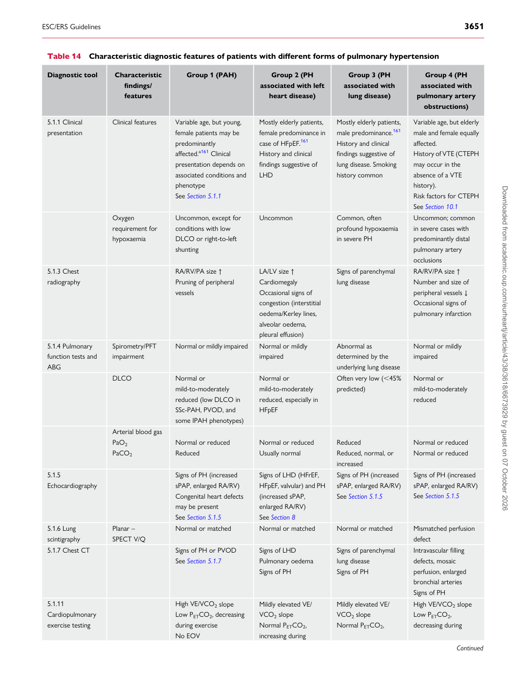
Table 14 Characteristic diagnostic features of patients with different forms of pulmonary hypertension

Diagnostic tool Characteristic

findings/ features Group 1 (PAH) Group 2 (PH

associated with left

heart disease) Group 3 (PH

associated with lung disease) Group 4 (PH

associated with pulmonary artery

obstructions)

5.1.1 Clinical presentation Clinical features Variable age, but young, female patients may be

predominantly affected.a161 Clinical presentation depends on associated conditions and phenotype See Section 5.1.1 Mostly elderly patients, female predominance in

case of HFpEF.161 History and clinical findings suggestive of LHD Mostly elderly patients, male predominance.161

History and clinical findings suggestive of lung disease. Smoking history common Variable age, but elderly male and female equally

affected. History of VTE (CTEPH may occur in the absence of a VTE history). Risk factors for CTEPH See Section 10.1

Oxygen requirement for hypoxaemia Uncommon, except for

conditions with low DLCO or right-to-left shunting Uncommon Common, often

profound hypoxaemia in severe PH Uncommon; common

in severe cases with predominantly distal pulmonary artery occlusions

5.1.3 Chest

radiography RA/RV/PA size ↑

Pruning of peripheral vessels LA/LV size ↑

Cardiomegaly Occasional signs of congestion (interstitial oedema/Kerley lines, alveolar oedema, pleural effusion) Signs of parenchymal

lung disease RA/RV/PA size ↑

Number and size of peripheral vessels ↓ Occasional signs of pulmonary infarction

5.1.4 Pulmonary function tests and

ABG Spirometry/PFT impairment Normal or mildly impaired Normal or mildly impaired Abnormal as determined by the

underlying lung disease Normal or mildly impaired

DLCO Normal or

mild-to-moderately reduced (low DLCO in SSc-PAH, PVOD, and some IPAH phenotypes) Normal or

mild-to-moderately reduced, especially in HFpEF Often very low (,45%

predicted) Normal or

mild-to-moderately reduced

Arterial blood gas PaO2 PaCO2 Normal or reduced

Reduced Normal or reduced

Usually normal Reduced

Reduced, normal, or increased Normal or reduced

Normal or reduced

5.1.5 Echocardiography Signs of PH (increased sPAP, enlarged RA/RV)

Congenital heart defects may be present See Section 5.1.5 Signs of LHD (HFrEF, HFpEF, valvular) and PH

(increased sPAP, enlarged RA/RV) See Section 8 Signs of PH (increased sPAP, enlarged RA/RV)

See Section 5.1.5 Signs of PH (increased sPAP, enlarged RA/RV)

See Section 5.1.5

5.1.6 Lung

scintigraphy Planar –

SPECT V/Q Normal or matched Normal or matched Normal or matched Mismatched perfusion

defect

5.1.7 Chest CT Signs of PH or PVOD

See Section 5.1.7 Signs of LHD

Pulmonary oedema Signs of PH Signs of parenchymal

lung disease Signs of PH Intravascular filling

defects, mosaic perfusion, enlarged bronchial arteries Signs of PH

5.1.11 Cardiopulmonary

exercise testing High VE/VCO2 slope

Low PETCO2, decreasing during exercise No EOV Mildly elevated VE/

VCO2 slope Normal PETCO2, increasing during Mildly elevated VE/

VCO2 slope Normal PETCO2, High VE/VCO2 slope

Low PETCO2, decreasing during

Continued Downloaded from academic.oup.com/eurheartj/article/43/38/3618/6673929 by guest on 07 October 2026