p.33 抽出テキスト
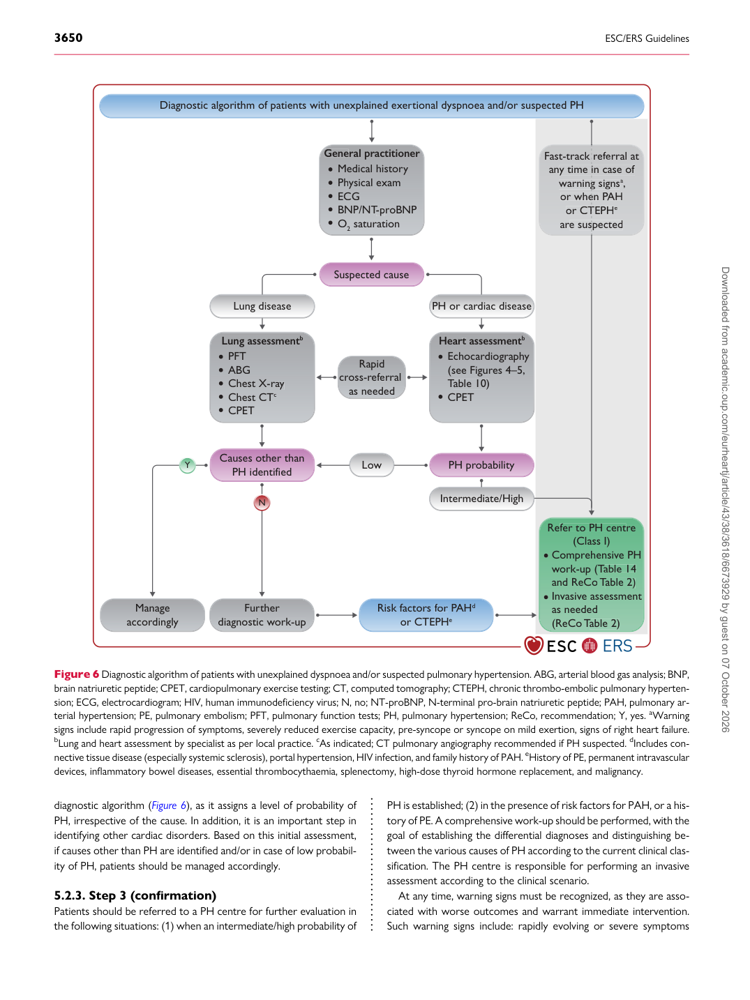
diagnostic algorithm (Figure 6), as it assigns a level of probability of PH, irrespective of the cause. In addition, it is an important step in identifying other cardiac disorders. Based on this initial assessment, if causes other than PH are identified and/or in case of low probability of PH, patients should be managed accordingly.

5.2.3 Step 3 (confirmation)

Patients should be referred to a PH centre for further evaluation in the following situations: (1) when an intermediate/high probability of PH is established; (2) in the presence of risk factors for PAH, or a history of PE. A comprehensive work-up should be performed, with the goal of establishing the differential diagnoses and distinguishing between the various causes of PH according to the current clinical classification. The PH centre is responsible for performing an invasive assessment according to the clinical scenario.

At any time, warning signs must be recognized, as they are associated with worse outcomes and warrant immediate intervention. Such warning signs include: rapidly evolving or severe symptoms

Diagnostic al g orithm of patients with unexplained e x e r tional dyspnoea and/or suspected PH

Suspected cause

PH p r obability

Causes other than

PH identified Medical histo r y

P h ysical exam ECG BNP/N T -p

T T r oBNP O 2 saturation

Gene G ra l prac titione r

N

PFT ABG Chest X-r a y

a a Chest CT c CPET

Lung ass e ss men t b

Echoca r diogr a p h y (see Figu r es 4–5, T able 10)

T

T CPET

He ar t

r r ass e ss men t b

Y Lung disease L o w PH or ca r diac disease

Intermediate/High Fast-track r e f e

f f r ral at

a n y time in case of

i warning signs

g a , or when

e P AH

P P or CTEPH

T e a r e suspected

s

Re f er to PH cent

f f r e

(Class I) Comp r ehensi v e PH w ork-up ( T able 14

T

T and ReCo T able 2)

T

T I n vasi v e assessment as needed (ReCo T able 2)

T

T

R a pid c r oss- r e f e

f f r ral as needed

Fu r ther diagnostic w ork-up Risk factors f or

f f P AH

P

P d

or CTEPH e

Manage acco r ding l y

Figure 6 Diagnostic algorithm of patients with unexplained dyspnoea and/or suspected pulmonary hypertension. ABG, arterial blood gas analysis; BNP, brain natriuretic peptide; CPET, cardiopulmonary exercise testing; CT, computed tomography; CTEPH, chronic thrombo-embolic pulmonary hypertension; ECG, electrocardiogram; HIV, human immunodeficiency virus; N, no; NT-proBNP, N-terminal pro-brain natriuretic peptide; PAH, pulmonary arterial hypertension; PE, pulmonary embolism; PFT, pulmonary function tests; PH, pulmonary hypertension; ReCo, recommendation; Y, yes. aWarning signs include rapid progression of symptoms, severely reduced exercise capacity, pre-syncope or syncope on mild exertion, signs of right heart failure. bLung and heart assessment by specialist as per local practice. cAs indicated; CT pulmonary angiography recommended if PH suspected. dIncludes connective tissue disease (especially systemic sclerosis), portal hypertension, HIV infection, and family history of PAH. eHistory of PE, permanent intravascular devices, inflammatory bowel diseases, essential thrombocythaemia, splenectomy, high-dose thyroid hormone replacement, and malignancy. Downloaded from academic.oup.com/eurheartj/article/43/38/3618/6673929 by guest on 07 October 2026