p.30 抽出テキスト
Incomplete assessments must be avoided, as this may lead to misdiagnosis. As a minimum, mixed venous oxygen saturation (SvO2) and arterial oxygen saturation (SaO2) should be determined. A stepwise assessment of oxygen saturation should be performed in patients with SvO2 .75% and whenever a left-to-right shunt is suspected. Cardiac output (CO) should be assessed by the direct Fick method or thermodilution (mean values of at least three measurements). The indirect Fick method is considered to be less reliable than thermodilution;127 however, thermodilution should not be used in the presence of shunts. Pulmonary vascular resistance ([mPAP−PAWP]/CO) should be calculated for each patient. All pressure measurements, including PAWP, should be performed at end expiration (without breath-holding manoeuvre). In patients with large intrathoracic pressure changes during the respiratory cycle (i.e. COPD, obesity, during exercise), it is appropriate to average over at least three to four respiratory cycles. If no reliable PAWP curve can be obtained, or if the PAWP values are implausible, additional measurement of LV end-diastolic pressure should be considered to avoid misclassification. Saturations taken with the catheter in the wedged position can confirm an accurate PAWP.128

5.1.12.2 Vasoreactivity testing

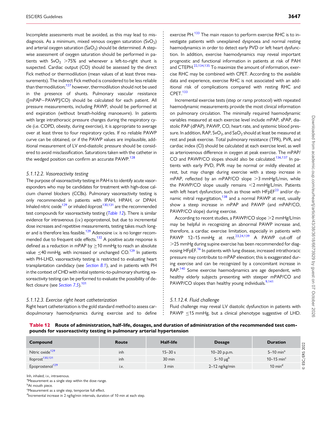
The purpose of vasoreactivity testing in PAH is to identify acute vasoresponders who may be candidates for treatment with high-dose calcium channel blockers (CCBs). Pulmonary vasoreactivity testing is only recommended in patients with IPAH, HPAH, or DPAH. Inhaled nitric oxide129 or inhaled iloprost130,131 are the recommended test compounds for vasoreactivity testing (Table 12). There is similar evidence for intravenous (i.v.) epoprostenol, but due to incremental dose increases and repetitive measurements, testing takes much longer and is therefore less feasible.129 Adenosine i.v. is no longer recommended due to frequent side effects.132 A positive acute response is defined as a reduction in mPAP by ≥10 mmHg to reach an absolute value ≤40 mmHg, with increased or unchanged CO.129 In patients with PH-LHD, vasoreactivity testing is restricted to evaluating heart transplantation candidacy (see Section 8.1), and in patients with PH in the context of CHD with initial systemic-to-pulmonary shunting, vasoreactivity testing can be performed to evaluate the possibility of defect closure (see Section 7.5).101

5.1.12.3 Exercise right heart catheterization

Right heart catheterization is the gold standard method to assess cardiopulmonary haemodynamics during exercise and to define exercise PH.133 The main reason to perform exercise RHC is to investigate patients with unexplained dyspnoea and normal resting haemodynamics in order to detect early PVD or left heart dysfunction. In addition, exercise haemodynamics may reveal important prognostic and functional information in patients at risk of PAH and CTEPH.22,134,135 To maximize the amount of information, exercise RHC may be combined with CPET. According to the available data and experience, exercise RHC is not associated with an additional risk of complications compared with resting RHC and CPET.133

Incremental exercise tests (step or ramp protocol) with repeated haemodynamic measurements provide the most clinical information on pulmonary circulation. The minimally required haemodynamic variables measured at each exercise level include mPAP, sPAP, diastolic PAP (dPAP), PAWP, CO, heart rate, and systemic blood pressure. In addition, RAP, SvO2, and SaO2 should at least be measured at rest and peak exercise. Total pulmonary resistance (TPR), PVR, and cardiac index (CI) should be calculated at each exercise level, as well as arteriovenous difference in oxygen at peak exercise. The mPAP/ CO and PAWP/CO slopes should also be calculated.136,137 In patients with early PVD, PVR may be normal or mildly elevated at rest, but may change during exercise with a steep increase in mPAP, reflected by an mPAP/CO slope .3 mmHg/L/min, while the PAWP/CO slope usually remains ,2 mmHg/L/min. Patients with left heart dysfunction, such as those with HFpEF23 and/or dynamic mitral regurgitation,138 and a normal PAWP at rest, usually show a steep increase in mPAP and PAWP (and mPAP/CO, PAWP/CO slope) during exercise. According to recent studies, a PAWP/CO slope .2 mmHg/L/min may be helpful in recognizing an abnormal PAWP increase and, therefore, a cardiac exercise limitation, especially in patients with PAWP 12–15 mmHg at rest.23,24,139 A PAWP cut-off of .25 mmHg during supine exercise has been recommended for diagnosing HFpEF.16 In patients with lung disease, increased intrathoracic pressure may contribute to mPAP elevation; this is exaggerated during exercise and can be recognized by a concomitant increase in RAP.140 Some exercise haemodynamics are age dependent, with healthy elderly subjects presenting with steeper mPAP/CO and PAWP/CO slopes than healthy young individuals.9,141

5.1.12.4 Fluid challenge

Fluid challenge may reveal LV diastolic dysfunction in patients with PAWP ≤15 mmHg, but a clinical phenotype suggestive of LHD.

Table 12 Route of administration, half-life, dosages, and duration of administration of the recommended test compounds for vasoreactivity testing in pulmonary arterial hypertension

Compound Route Half-life Dosage Duration Nitric oxide129 inh 15–30 s 10–20 p.p.m. 5–10 mina Iloprost130,131 inh 30 min 5–10 μgb 10–15 minc Epoprostenol129 i.v. 3 min 2–12 ng/kg/min 10 mind © ESC/ERS 2022 Inh, inhaled; i.v., intravenous. aMeasurement as a single step within the dose range. bAt mouth piece. cMeasurement as a single step, temporize full effect. dIncremental increase in 2 ng/kg/min intervals, duration of 10 min at each step. Downloaded from academic.oup.com/eurheartj/article/43/38/3618/6673929 by guest on 07 October 2026