p.29 抽出テキスト
intracardiac shunt, and retrograde flow. By combining contrast magnetic resonance (MR) angiography and pulmonary perfusion imaging with late gadolinium-enhancement imaging of the myocardium, a complete picture of the heart and pulmonary vasculature can be obtained (see Supplementary Data, Table S2 for cMRI indices and normal values). A limitation is that there is no established method with which to estimate PAP. Even though the cost and availability of the technique precludes its use in the early diagnosis of PAH, it is sensitive in detecting early signs of PH and diagnosing CHD.117

5.1.9 Blood tests and immunology

The initial diagnostic assessment of patients with newly diagnosed PH/PAH aims to identify comorbidities and possible causes or complications of PH. Laboratory tests that should be obtained at the time of PH diagnosis include: blood counts (including haemoglobin [Hb]); serum electrolytes (sodium, potassium); kidney function (creatinine, calculation of estimated glomerular filtration rate, and urea); uric acid; liver parameters (alanine aminotransferase, aspartate aminotransferase, and alkaline phosphatase, γ-glutamyl transpeptidase, bilirubin); iron status (serum iron, transferrin saturation, and ferritin); and BNP or NT-proBNP. In addition, serological studies should include testing for hepatitis viruses and HIV. Basic immunology laboratory work-up is recommended, including screening tests for anti-nuclear antibodies, anti-centromere antibodies, and anti-Ro. Screening for biological markers of antiphospholipid syndrome is recommended in patients with CTEPH. Additional thrombophilia screening is not generally recommended, unless therapeutic consequences are to be expected.118 Pulmonary arterial hypertension and other forms of severe PH can be associated with thyroid function disorders; hence, laboratory screening should include at least thyroid-stimulating hormone.

5.1.10 Abdominal ultrasound

An abdominal ultrasound examination should be part of the comprehensive diagnostic work-up of patients with newly diagnosed PH, particularly if liver disease is suspected. A major objective is to search for liver disease and/or portal hypertension, or portocaval shunt (Abernethy malformation). During the course of the disease, patients with PH may develop secondary organ dysfunction mainly affecting the liver and kidneys.119 In these patients, abdominal ultrasound is needed for differential diagnostic reasons and to assess the extent of organ damage.

5.1.11 Cardiopulmonary exercise testing

Cardiopulmonary exercise testing (CPET) is a useful tool to assess the underlying pathophysiologic mechanisms leading to exercise intolerance. Patients with PAH show a typical pattern, with a low endtidal partial pressure of carbon dioxide (PETCO2), high ventilatory equivalent for carbon dioxide (VE/VCO2), low oxygen pulse (VO2/ HR), and low peak oxygen uptake (VO2).120 These findings should prompt consideration of PVD. In patients with LHD or COPD, such a pattern may indicate an additional pulmonary vascular limitation.121,122 In populations at risk of PAH, such as those with SSc, a normal peak VO2 seems to exclude the diagnosis of PAH.123 5.1.12. Right heart catheterization, vasoreactivity, exercise, and fluid challenge 5.1.12.1. Right heart catheterization Right heart catheterization is the gold standard for diagnosing and classifying PH. Performing RHC requires expertise and meticulous methodology following standardized protocols. In addition to diagnosing and classifying PH, clinical indications include haemodynamic assessment of heart or LTx candidates124 and evaluating congenital cardiac shunts. Interpreting invasive haemodynamics should be done in the context of the clinical picture and other diagnostic investigations. When performed in PH centres, the frequencies of serious adverse events (1.1%) and procedure-related mortality (0.055%) are low.125 A known thrombus or tumour in the RV or RA, recently implanted (,1 month) pacemaker, mechanical right heart valve, TriClip, and an acute infection are contraindications to RHC; the risk:benefit ratio should be individually assessed before each examination and discussed with the patient. The most feared complication of RHC is perforation of a PA.

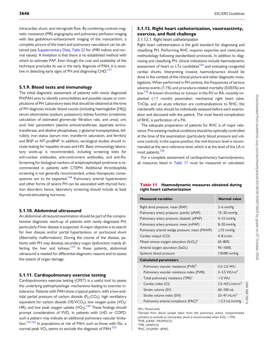
The adequate preparation of patients for RHC is of major relevance. Pre-existing medical conditions should be optimally controlled at the time of the examination (particularly blood pressure and volume control). In the supine position, the mid-thoracic level is recommended as the zero reference level, which is at the level of the LA in most patients.126 For a complete assessment of cardiopulmonary haemodynamics, all measures listed in Table 11 must be measured or calculated.

Table 11 Haemodynamic measures obtained during right heart catheterization

Measured variables Normal value

Right atrial pressure, mean (RAP) 2–6 mmHg Pulmonary artery pressure, systolic (sPAP) 15–30 mmHg Pulmonary artery pressure, diastolic (dPAP) 4–12 mmHg Pulmonary artery pressure, mean (mPAP) 8–20 mmHg Pulmonary arterial wedge pressure, mean (PAWP) ≤15 mmHg Cardiac output (CO) 4–8 L/min Mixed venous oxygen saturation (SvO2)a 65–80% Arterial oxygen saturation (SaO2) 95–100% Systemic blood pressure 120/80 mmHg Calculated parameters

Pulmonary vascular resistance (PVR)b 0.3–2.0 WU Pulmonary vascular resistance index (PVRI) 3–3.5 WU·m2 Total pulmonary resistance (TPR)c ,3 WU Cardiac index (CI) 2.5–4.0 L/min·m2 Stroke volume (SV) 60–100 mL Stroke volume index (SVI) 33–47 mL/m2 Pulmonary arterial compliance (PAC)d .2.3 mL/mmHg © ESC/ERS 2022 WU, Wood units. aDerived from blood sample taken from the pulmonary artery; compartmental oximetry to exclude an intracardiac shunt is recommended when SvO2 .75%. bPVR, (mPAP−PAWP)/CO. cTPR, mPAP/CO. dPAC, SV/(sPAP−dPAP). Downloaded from academic.oup.com/eurheartj/article/43/38/3618/6673929 by guest on 07 October 2026