p.28 抽出テキスト
In patients presenting with a clinical picture of acute PE, chest CT may be helpful in detecting signs of hitherto undetected CTEPH, which may include the presence of the above CTEPH signs, and RV hypertrophy as a sign for chronicity.111,112 Detecting ‘acute on chronic’ PE is important, as it may impact the management of patients with presumed acute PE. Dual-energy CT (DECT) angiography and iodine subtraction mapping may provide additional diagnostic information by creating iodine maps,113 which reflect lung perfusion, thereby possibly increasing the diagnostic accuracy for CTEPH.114 Although increasingly used, the diagnostic value of DECT in the work-up of patients with PH has not been established. Digital subtraction angiography (DSA) is mainly used to confirm the diagnosis of CTEPH and to assess treatment options (i.e. operability or accessibility for BPA). Most centres use conventional two- or three-planar DSA. However, C-arm CT imaging may provide a higher spatial resolution, potentially identifying more target vessels for BPA and providing procedural guidance.115,116

5.1.8 Cardiac magnetic resonance imaging

Cardiac magnetic resonance imaging accurately and reproducibly assesses atrial and ventricular size, morphology, and function. Additional information on RV/LV myocardial strain can be obtained by applying tagging or by post-processing feature tracking. In addition, cMRI can be used to measure blood flow in the PA, aorta, and vena cava, allowing for quantifying stroke volume (SV),

Table 10 Additional echocardiographic signs suggestive of pulmonary hypertension

A: The ventricles B: Pulmonary

artery C: Inferior vena

cava and RA

RV/LV basal diameter/ area ratio .1.0 RVOT AT ,105 ms and/or

mid-systolic notching IVC diameter .21 mm with

decreased inspiratory collapse (,50% with a sniff or ,20% with quiet inspiration)

Flattening of the interventricular septum (LVEI .1.1 in systole and/or diastole) Early diastolic

pulmonary regurgitation velocity .2.2 m/s RA area (end-systole)

.18 cm2

TAPSE/sPAP ratio ,0.55 mm/mmHg PA diameter .AR diameter

PA diameter .25 mm © ESC/ERS 2022

AR, aortic root; IVC, inferior vena cava; LV, left ventricle; LVEI, left ventricle eccentricity index; PA, pulmonary artery; RA, right atrium; RV, right ventricle; RVOT AT, right ventricular outflow tract acceleration time; sPAP, systolic pulmonary arterial pressure; TAPSE, tricuspid annular plane systolic excursion; TRV, tricuspid regurgitation velocity. aSigns contributing to assessing the probability of PH in addition to TRV (see Figure 5). Signs from at least two categories (A/B/C) must be present to alter the level of echocardiographic probability of PH.

P eak tricuspid r egurgitation v elocity (m/s)

P r esence of other echo PH signs b (see T able 10)

T

T

N N

≤ 2.8 a 2.9–3.4

PH P R O

R R B ABILITY INTERMEDI A TE

A A PH P R O

R R B ABILITY HIGH

PH P R O

R R B ABILITY L O W >3.4

Y Y Y es/No

Y

Y

Risk factors or associated conditions

o f or

f f P AH or CTEPH

P P

N N

Echo f oll

f f o w-up (Class IIa) Fu r ther inte r v ention c /

RHC (if indicated d )

(Class I)

Alternati v e diagnosis

(Class IIa) Y Y Y es/No

Y Y

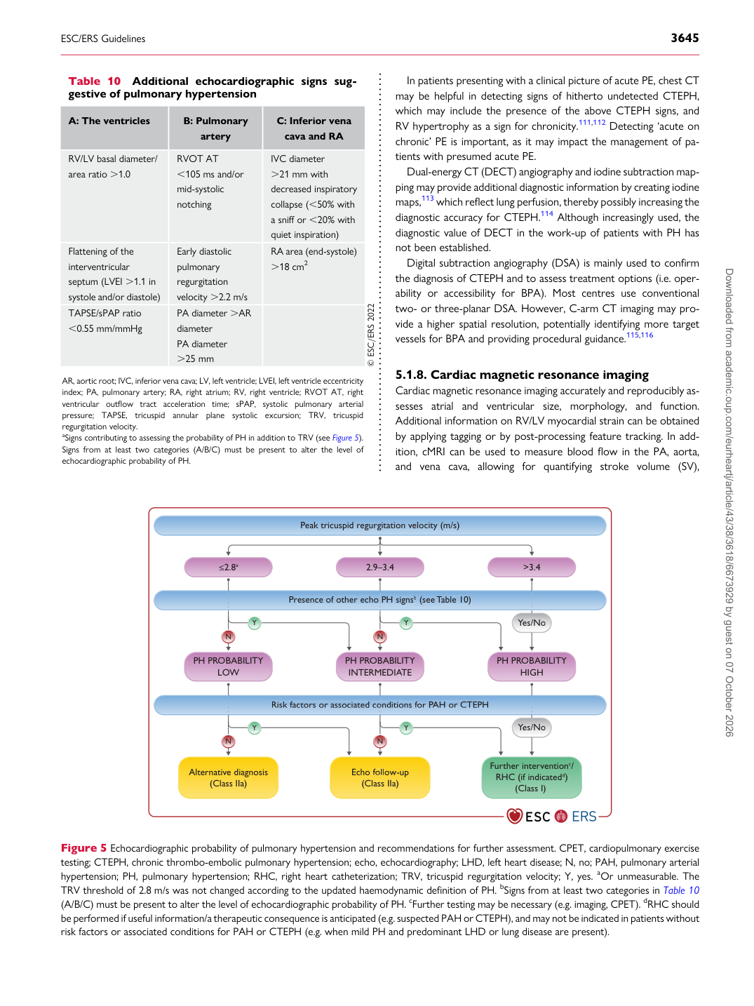
Figure 5 Echocardiographic probability of pulmonary hypertension and recommendations for further assessment. CPET, cardiopulmonary exercise testing; CTEPH, chronic thrombo-embolic pulmonary hypertension; echo, echocardiography; LHD, left heart disease; N, no; PAH, pulmonary arterial hypertension; PH, pulmonary hypertension; RHC, right heart catheterization; TRV, tricuspid regurgitation velocity; Y, yes. aOr unmeasurable. The TRV threshold of 2.8 m/s was not changed according to the updated haemodynamic definition of PH. bSigns from at least two categories in Table 10 (A/B/C) must be present to alter the level of echocardiographic probability of PH. cFurther testing may be necessary (e.g. imaging, CPET). dRHC should be performed if useful information/a therapeutic consequence is anticipated (e.g. suspected PAH or CTEPH), and may not be indicated in patients without risk factors or associated conditions for PAH or CTEPH (e.g. when mild PH and predominant LHD or lung disease are present). Downloaded from academic.oup.com/eurheartj/article/43/38/3618/6673929 by guest on 07 October 2026