p.27 抽出テキスト
CTEPH may be present in 7–10% of patients with PVOD/PCH or PAH.106,107 Deposition of the perfusion agent in extrapulmonary organs may hint to cardiac or pulmonary right-to-left shunting and has been reported in CHD, hepato-pulmonary syndrome, and pulmonary arteriovenous malformations (PAVMs).68

5.1.7 Non-contrast and contrast-enhanced chest

computed tomography examinations, and digital subtraction angiography Computed tomography (CT) imaging may provide important information for patients with unexplained dyspnoea or suspected/confirmed PH. The CT signs suggesting the presence of PH include an enlarged PA diameter, a PA-to-aorta ratio .0.9, and enlarged right heart chambers.68 A combination of three parameters (PA diameter ≥30 mm, RVOT wall thickness ≥6 mm, and septal deviation ≥140° [or RV:LV ratio ≥1]) is highly predictive of PH.108 Non-contrast chest CT can help determine the cause of PH when there are features of parenchymal lung disease, and may also point towards the presence of PVOD/PCH by showing centrilobular ground-glass opacities (which may also be found in PAH), septal lines, and lymphadenopathy.68

Computed tomography pulmonary angiography (CTPA) is mainly used to detect direct or indirect signs of CTEPH, such as filling defects (including thrombus adhering to the vascular wall), webs or bands in the PAs, PA retraction/dilatation, mosaic perfusion, and enlarged bronchial arteries. Importantly, the diagnostic accuracy of CTPA for CTEPH is limited (at the patient level, sensitivity and specificity are 76% and 96%, respectively),109 but was reported to be higher when modern, high-quality multi-detector CT scanners were used and when interpreted by experienced readers.109,110 Computed tomography pulmonary angiography may also be used to detect other cardiovascular abnormalities, including intracardiac shunts, abnormal pulmonary venous return, patent ductus arteriosus, and PAVMs.

RV Ao LA LV

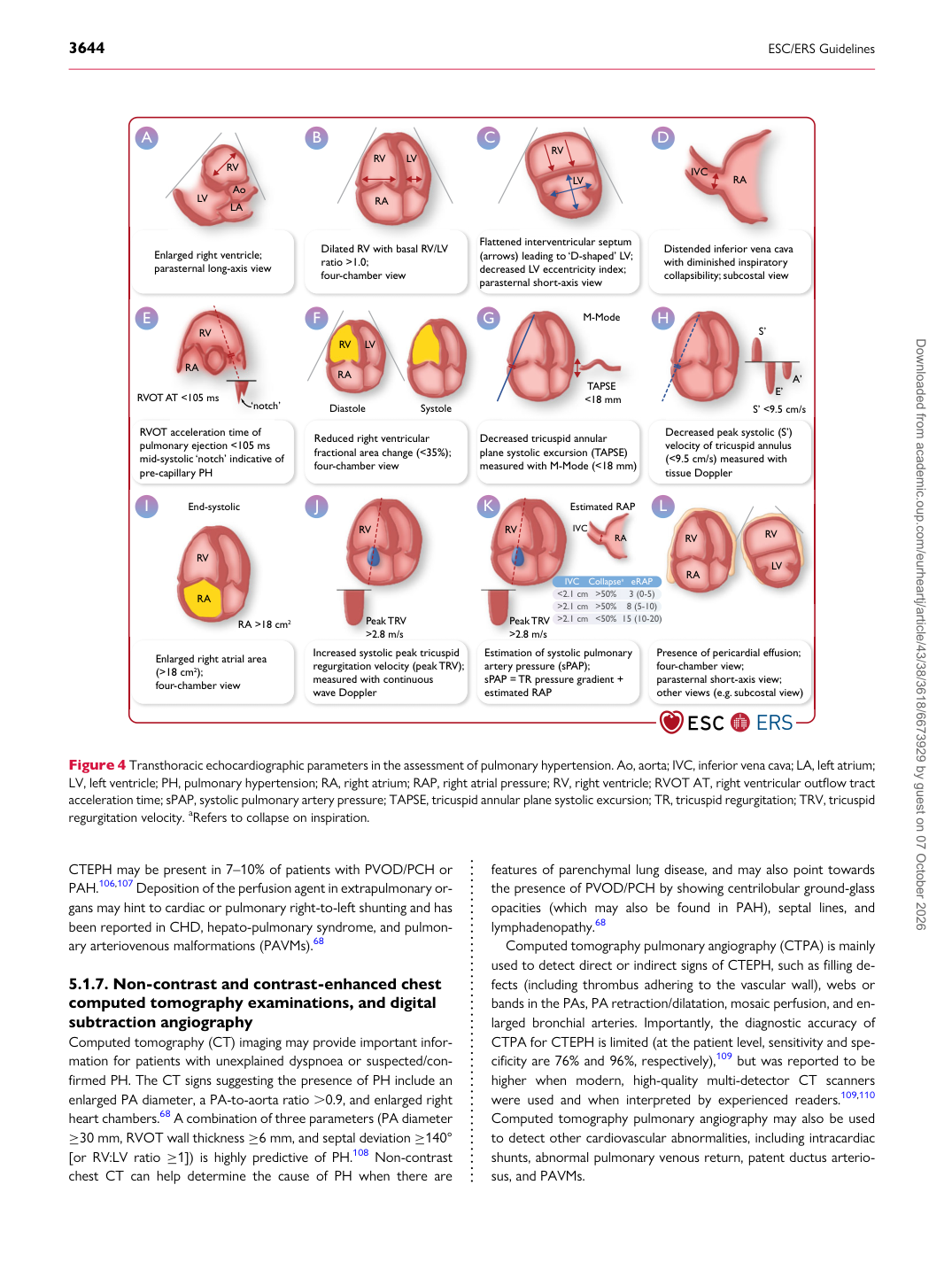
Enlarged right ventricle; parasternal long-axis view

A

RV

RA LV

Dilated RV with basal RV/LV ratio >1.0; four-chamber view

B

RV

LV

Flattened interventricular septum (arrows) leading to ‘D-shaped’ LV; decreased LV eccentricity index; parasternal short-axis view

C

RA IVC

Distended inferior vena cava with diminished inspiratory collapsibility; subcostal view

D

RVOT acceleration time of pulmonary ejection <105 ms mid-systolic ‘notch’ indicative of pre-capillary PH

E

Reduced right ventricular fractional area change (<35%); four-chamber view

F RV

RA

RVOT AT <105 ms

‘notch’ Diastole Systole

RV

RA LV

TAPSE <18 mm

M-Mode

Decreased tricuspid annular plane systolic excursion (TAPSE) measured with M-Mode (<18 mm)

G

Decreased peak systolic (S’) velocity of tricuspid annulus (<9.5 cm/s) measured with tissue Doppler

H

Enlarged right atrial area (>18 cm2); four-chamber view

I E’

S’

S’ <9.5 cm/s

RV

RA A’

RV

Increased systolic peak tricuspid regurgitation velocity (peak TRV); measured with continuous wave Doppler

J

Estimation of systolic pulmonary artery pressure (sPAP); sPAP = TR pressure gradient + estimated RAP

K

Presence of pericardial effusion; four-chamber view; parasternal short-axis view; other views (e.g. subcostal view)

L

RV

RA

Estimated RAP

RV RA IVC

IVC Collapsea eRAP <2.1 cm >50% 3 (0-5)

Peak TRV >2.8 m/s >2.1 cm >50% 8 (5-10) >2.1 cm <50% 15 (10-20) RV LV

Peak TRV >2.8 m/s

End-systolic

RA >18 cm2

Figure 4 Transthoracic echocardiographic parameters in the assessment of pulmonary hypertension. Ao, aorta; IVC, inferior vena cava; LA, left atrium; LV, left ventricle; PH, pulmonary hypertension; RA, right atrium; RAP, right atrial pressure; RV, right ventricle; RVOT AT, right ventricular outflow tract acceleration time; sPAP, systolic pulmonary artery pressure; TAPSE, tricuspid annular plane systolic excursion; TR, tricuspid regurgitation; TRV, tricuspid regurgitation velocity. aRefers to collapse on inspiration. Downloaded from academic.oup.com/eurheartj/article/43/38/3618/6673929 by guest on 07 October 2026