p.25 抽出テキスト
at diagnosis and follow-up is common in PAH and associated with unfavourable outcomes.80 Elevated PaCO2 is very unusual in PAH and reflects alveolar hypoventilation, which in itself may be a cause of PH. Overnight oximetry or polysomnography should be performed if there is suspicion of sleep-disordered breathing or hypoventilation.81

5.1.5 Echocardiography

Independent of the underlying aetiology, PH leads to RV pressure overload and dysfunction, which can be detected by echocardiography.82–84 When performed accurately, echocardiography provides comprehensive information on right and left heart morphology, RV and LV function, and valvular abnormalities, and gives estimates of haemodynamic parameters. Echocardiography is also a valuable tool with which to detect the cause of suspected or confirmed PH, particularly with respect to PH associated with LHD or CHD. Yet, echocardiography alone is insufficient to confirm a diagnosis of PH, which requires RHC.

Given the heterogeneous nature of PH and the peculiar geometry of the RV, there is no single echocardiographic parameter that reliably informs about PH status and underlying aetiology. Therefore, a comprehensive echocardiographic evaluation for suspected PH includes estimating the systolic pulmonary arterial pressure (sPAP) and detecting additional signs suggestive of PH, aiming at assigning an echocardiographic level of probability of PH. Echocardiographic findings of PH, including estimating pressure and signs of RV overload and/or dysfunction, are summarized in Figure 4.

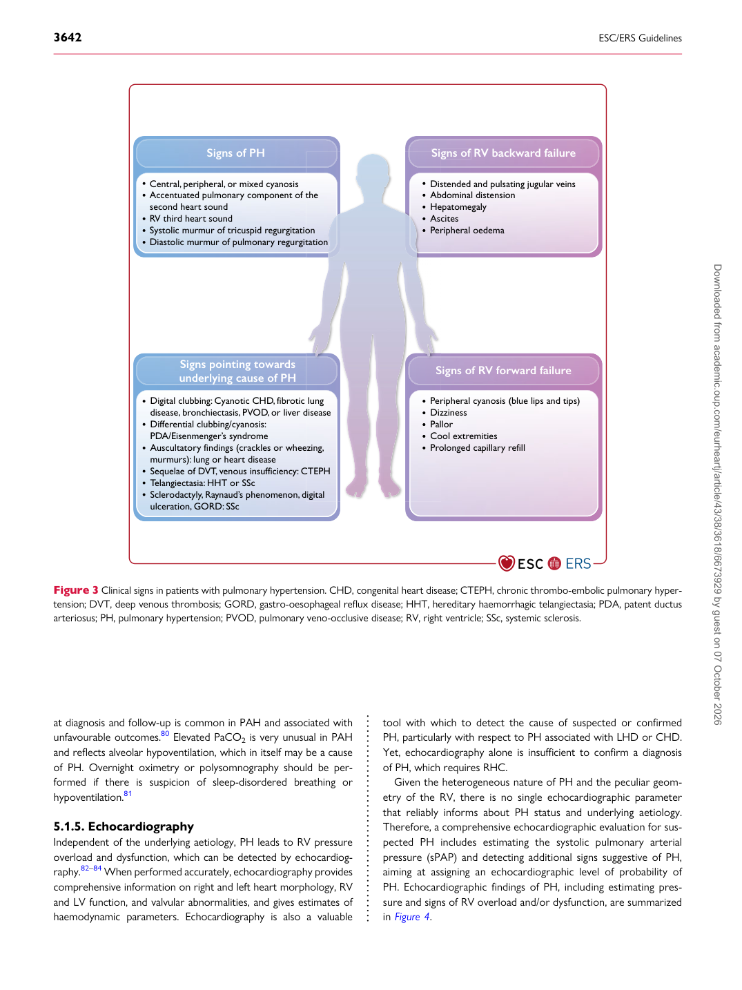
Signs of PH

Central, peripheral, or mixed cyanosis Accentuated pulmonary component of the second heart sound RV third heart sound Systolic murmur of tricuspid regurgitation Diastolic murmur of pulmonary regurgitation

Signs pointing towards underlying cause of PH Digital clubbing: Cyanotic CHD, fibrotic lung disease, bronchiectasis, PVOD, or liver disease Differential clubbing/cyanosis: PDA/Eisenmenger’s syndrome Auscultatory findings (crackles or wheezing, murmurs): lung or heart disease Sequelae of DVT, venous insufficiency: CTEPH Telangiectasia: HHT or SSc Sclerodactyly, Raynaud’s phenomenon, digital ulceration, GORD: SSc Signs of RV backward failure

Signs of RV forward failure

Distended and pulsating jugular veins Abdominal distension Hepatomegaly Ascites Peripheral oedema

Peripheral cyanosis (blue lips and tips) Dizziness Pallor Cool extremities Prolonged capillary refill

the

on tation

lung disease

ezing,

CTEPH

digital Sig

S

Dis Ab He Asc P er

P er Diz Pal Co P r o

Figure 3 Clinical signs in patients with pulmonary hypertension. CHD, congenital heart disease; CTEPH, chronic thrombo-embolic pulmonary hypertension; DVT, deep venous thrombosis; GORD, gastro-oesophageal reflux disease; HHT, hereditary haemorrhagic telangiectasia; PDA, patent ductus arteriosus; PH, pulmonary hypertension; PVOD, pulmonary veno-occlusive disease; RV, right ventricle; SSc, systemic sclerosis. Downloaded from academic.oup.com/eurheartj/article/43/38/3618/6673929 by guest on 07 October 2026