p.24 抽出テキスト
5.1.3 Chest radiography

Chest radiography presents abnormal findings in most patients with PH; however, a normal chest X-ray does not exclude PH.68 Radiographic signs of PH include a characteristic configuration of the cardiac silhouette due to right heart (right atrium [RA]/RV) and PA enlargement, sometimes with pruning of the peripheral vessels. In addition, signs of the underlying cause of PH, such as LHD or lung disease, may be found (Table 9).25,26,60,69,70

5.1.4 Pulmonary function tests and arterial blood

gases Pulmonary function tests (PFTs) and analysis of arterial blood gas (ABG) or arterialized capillary blood are necessary to distinguish between PH groups, assess comorbidities and the need for supplementary oxygen, and determine disease severity. The initial work-up of patients with suspected PH should comprise forced spirometry, body plethysmography, lung diffusion capacity for carbon monoxide (DLCO), and ABG. In patients with PAH, PFTs are usually normal or may show mild restrictive, obstructive, or combined abnormalities.71,72 More severe PFT abnormalities are occasionally found in patients with PAH associated with CHD,73 and those with group 3 PH. The DLCO may be normal in patients with PAH, although it is usually mildly reduced.71 A severely reduced DLCO (,45% of the predicted value) in the presence of otherwise normal PFTs can be found in PAH associated with systemic sclerosis (SSc), PVOD, in PH group 3—associated with emphysema, interstitial lung disease (ILD), or combined pulmonary fibrosis and emphysema—and in some PAH phenotypes.74 A low DLCO is associated with a poor prognosis in several forms of PH.75–78

Patients with PAH usually have normal or slightly reduced partial pressure of arterial oxygen (PaO2). Severe reduction of PaO2 might raise suspicion for patent foramen ovale, hepatic disease, other abnormalities with right-to-left shunt (e.g. septal defect), or low-DLCO-associated conditions. Partial pressure of arterial carbon dioxide (PaCO2) is typically lower than normal due to alveolar hyperventilation.79 Low PaCO2

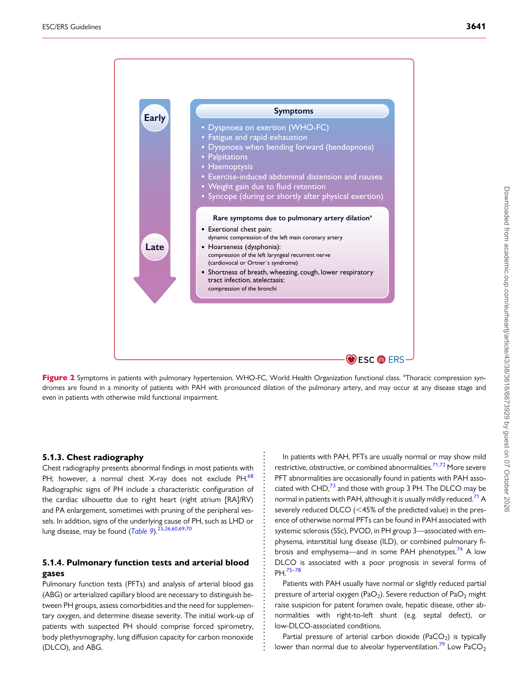
Symptoms

Dyspnoea on exertion (WHO-FC) Fatigue and rapid exhaustion Dyspnoea when bending forward (bendopnoea) Palpitations Haemoptysis Exercise-induced abdominal distension and nausea Weight gain due to fluid retention Syncope (during or shortly after physical exertion)

Rare symptoms due to pulmonary artery dilationa Exertional chest pain: dynamic compression of the left main coronary artery

Early

Late Hoarseness (dysphonia): compression of the left laryngeal recurrent nerve (cardiovocal or Ortner ́s syndrome) Shortness of breath, wheezing, cough, lower respiratory tract infection, atelectasis: compression of the bronchi

Figure 2 Symptoms in patients with pulmonary hypertension. WHO-FC, World Health Organization functional class. aThoracic compression syndromes are found in a minority of patients with PAH with pronounced dilation of the pulmonary artery, and may occur at any disease stage and even in patients with otherwise mild functional impairment. Downloaded from academic.oup.com/eurheartj/article/43/38/3618/6673929 by guest on 07 October 2026