p.27 抽出テキスト
the association of diastolic dysfunction with post-operative adverse events, including pulmonary oedema, AF, and mortality.134–138 A meta-analysis including 3876 patients undergoing NCS found preoperative diastolic dysfunction to be an independent risk factor for pulmonary oedema, congestive HF, and MI after surgery.139 However, a recent retrospective study, including 7312 patients, showed no association between the degree of diastolic dysfunction and in-hospital mortality or hospital length of stay in NCS patients.140 Awareness of diastolic dysfunction or high LV filling pressure (e.g. pulmonary hypertension, left atrial volume, E/e′ ratio) seems necessary to optimize peri-operative patient management; however, evidence does not support screening for diastolic dysfunction.

4.5.2 Stress tests

4.5.2.1. Exercise stress test Physical exercise, using a treadmill or bicycle ergometer, provides an estimate of functional capacity, evaluates blood pressure (BP) and heart rate response, and detects myocardial ischaemia through pathological ST-segment changes with poor sensitivity (61–73%) and specificity (60–80%).146 An exercise stress test alone should only be considered a valuable alternative to diagnose obstructive CAD if non-invasive imaging tests are unavailable.146 An exercise stress test is of no diagnostic value in patients with pre-existing ST-segment abnormalities (i.e. left bundle branch block, paced rhythm, Wolff–Parkinson–White [WPW] syndrome, ≥0.1 mV ST-segment depression on resting ECG, or taking digitalis). In addition, an exercise test is unsuitable for patients with limited exercise capacity, owing to their inability to reach their target heart rate. Therefore, an exercise stress test alone should only be considered a valuable alternative to diagnose obstructive CAD if non-invasive imaging tests are unavailable, or for assessing functional capacity when clinical history is ambiguous.146

4.5.2.2 Stress imaging

The use of stress imaging is appropriate for risk assessment in patients with clinical risk factors and poor functional capacity.147,148 The choice of the test is driven by local expertise. Selection, optimal, and safe performance of stress imaging should comply with related guidelines and recommendations.146,148,149 Stress imaging is not recommended in patients undergoing urgent surgery or with an unstable clinical condition. Evidence on the role of stress imaging for peri-operative risk prediction and patient management is largely based on inducible ischaemia by pharmacological stress testing, although no evidence indicates the superiority of pharmacological stress to exercise stress imaging in patients who are able to perform an adequate level of physical exercise. Several studies and meta-analyses have consistently defined clinical utility of pharmacological stress imaging for peri-operative risk assessment in patients undergoing NCS.150–154 Although RCTs related to post-operative outcome are lacking, there are large-scale prospective studies showing a risk-adjusted association of stress imaging results with perioperative cardiac complications.155–157 Studies and meta-analyses have demonstrated similar prognostic value of stress echocardiography and myocardial perfusion imaging for peri-operative risk assessment with slightly higher negative predictive value of stress echocardiography,152 but the overall accuracy varies with ischaemic heart disease (IHD) prevalence.151 A moderate-to-large perfusion defect on either test is highly sensitive for post-operative cardiac events.152,158 Normal stress imaging exams without resting abnormalities have high negative predictive value.159,160 However, positive predictive value of stress imaging for peri-operative cardiac events is relatively low and requires confirmation by other tests.150,152,161 In a recent retrospective study including 4494 patients, dobutamine stress echocardiography (DSE) provided modest incremental predictive value for peri-operative CV complications over clinical variables and was found to be useful as part of a stepwise approach in the risk stratification of patients undergoing intermediate- to highrisk NCS.157 The strongest predictors of post-operative adverse events determined so far are significant ischaemia (more than four ventricular segments) during DSE, ischaemic threshold (60% of agepredicted maximal heart rate), and a history of congestive HF.162,163 A negative DSE without resting wall motion abnormality has excellent negative predictive value, even when target heart rate cannot be achieved despite an aggressive DSE regimen.164 In asymptomatic patients if functional capacity is unknown, stress echocardiography also enables dynamic evaluation of LV systolic and diastolic function; valvular diseases such as aortic valve stenosis (AS), mitral valve stenosis (MS), and hypertrophic obstructive cardiomyopathy; and pulmonary hypertension 165 However, the role of DSE for risk

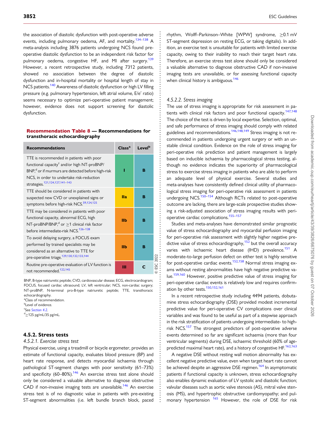
Recommendation Table 8 — Recommendations for transthoracic echocardiography

Recommendations Classa Levelb

TTE is recommended in patients with poor functional capacityc and/or high NT-proBNP/ BNP,d or if murmurs are detected before high-risk NCS, in order to undertake risk-reduction strategies.121,124,127,141–143 I B TTE should be considered in patients with suspected new CVD or unexplained signs or symptoms before high-risk NCS.59,124,125 IIa B TTE may be considered in patients with poor functional capacity, abnormal ECG, high NT-proBNP/BNP,d or ≥1 clinical risk factor before intermediate-risk NCS.126–128 IIb B To avoid delaying surgery, a FOCUS exam performed by trained specialists may be considered as an alternative to TTE for pre-operative triage.129,130,132,133,144 IIb B Routine pre-operative evaluation of LV function is not recommended.122,145 III C © ESC 2022

BNP, B-type natriuretic peptide; CVD, cardiovascular disease; ECG, electrocardiogram; FOCUS, focused cardiac ultrasound; LV, left ventricular; NCS, non-cardiac surgery; NT-proBNP, N-terminal pro-B-type natriuretic peptide; TTE, transthoracic echocardiography. aClass of recommendation. bLevel of evidence. cSee Section 4.2. d≥125 pg/mL/35 pg/mL. Downloaded from academic.oup.com/eurheartj/article/43/39/3826/6675076 by guest on 07 October 2026