p.93 抽出テキスト
CTb Echocardiographya PETd CMRc

Secondary tumour

(metastasis)f Benign primary

tumour Malignant primary

tumour

Diagnostic algorithm for cardiac masses

Non-invasive assessment of single or multiple cardiac masses

Tumour

Type Thrombus, vegetation, structural

Consider cardiac mass biopsy if malignant primary tumour suspected or diagnosis uncertain on imaginge

Lymphoma Sarcoma Angioma, lipoma, papillary fibroelastoma Myxoma

Leukaemias Lymphomas Melanomas Solid tumours: breast, lung, and oesophageal cancers

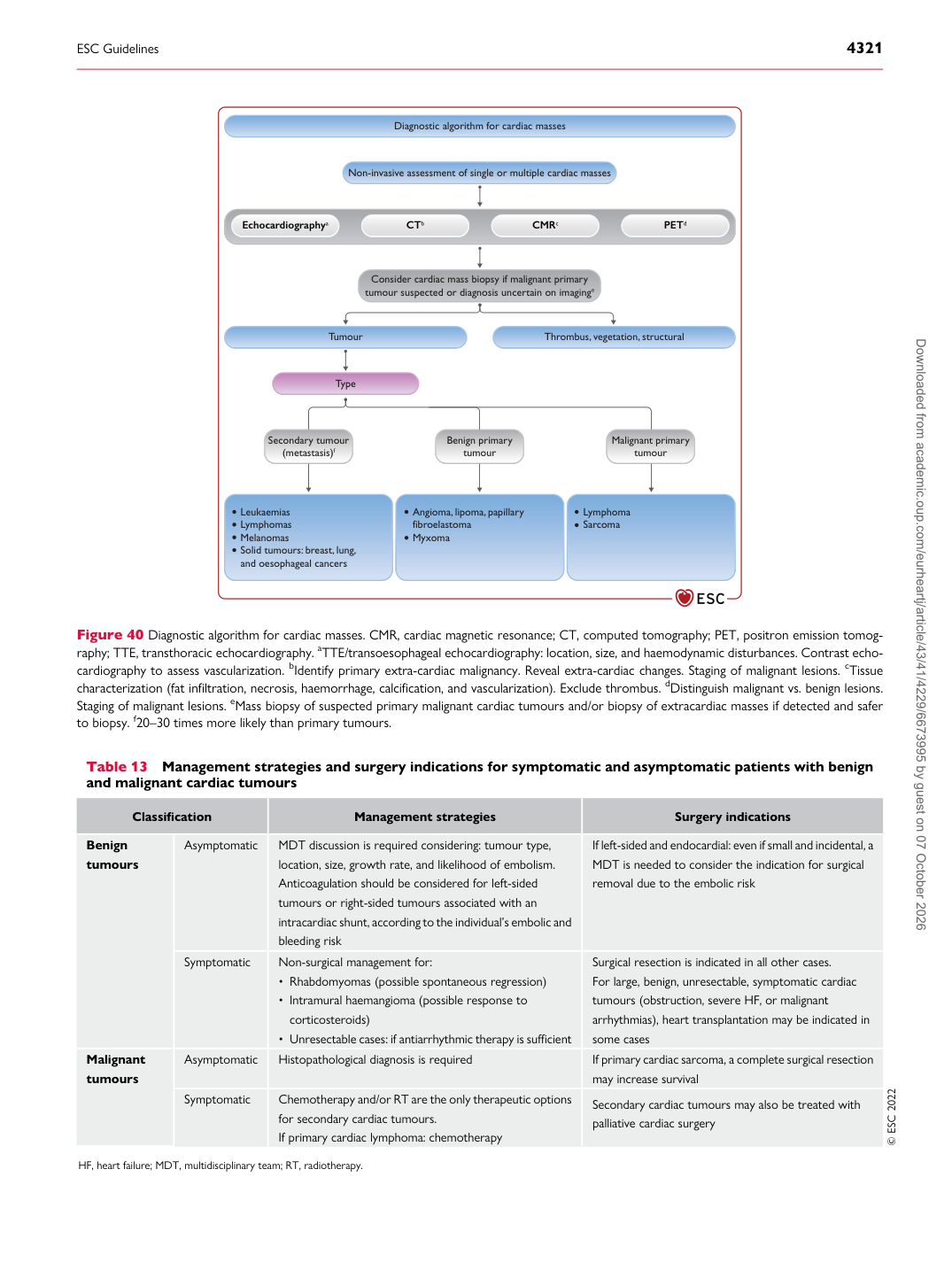
Figure 40 Diagnostic algorithm for cardiac masses. CMR, cardiac magnetic resonance; CT, computed tomography; PET, positron emission tomography; TTE, transthoracic echocardiography. aTTE/transoesophageal echocardiography: location, size, and haemodynamic disturbances. Contrast echocardiography to assess vascularization. bIdentify primary extra-cardiac malignancy. Reveal extra-cardiac changes. Staging of malignant lesions. cTissue characterization (fat infiltration, necrosis, haemorrhage, calcification, and vascularization). Exclude thrombus. dDistinguish malignant vs. benign lesions. Staging of malignant lesions. eMass biopsy of suspected primary malignant cardiac tumours and/or biopsy of extracardiac masses if detected and safer to biopsy. f20–30 times more likely than primary tumours.

Table 13 Management strategies and surgery indications for symptomatic and asymptomatic patients with benign and malignant cardiac tumours

Classification Management strategies Surgery indications

Benign tumours Asymptomatic MDT discussion is required considering: tumour type, location, size, growth rate, and likelihood of embolism.

Anticoagulation should be considered for left-sided tumours or right-sided tumours associated with an intracardiac shunt, according to the individual’s embolic and bleeding risk If left-sided and endocardial: even if small and incidental, a MDT is needed to consider the indication for surgical

removal due to the embolic risk

Symptomatic Non-surgical management for:

• Rhabdomyomas (possible spontaneous regression) • Intramural haemangioma (possible response to corticosteroids) • Unresectable cases: if antiarrhythmic therapy is sufficient Surgical resection is indicated in all other cases.

For large, benign, unresectable, symptomatic cardiac tumours (obstruction, severe HF, or malignant arrhythmias), heart transplantation may be indicated in some cases

Malignant tumours Asymptomatic Histopathological diagnosis is required If primary cardiac sarcoma, a complete surgical resection

may increase survival

Symptomatic Chemotherapy and/or RT are the only therapeutic options

for secondary cardiac tumours. If primary cardiac lymphoma: chemotherapy Secondary cardiac tumours may also be treated with

palliative cardiac surgery © ESC 2022

HF, heart failure; MDT, multidisciplinary team; RT, radiotherapy. Downloaded from academic.oup.com/eurheartj/article/43/41/4229/6673995 by guest on 07 October 2026