p.88 抽出テキスト
8.3 Coronary artery disease

Any vascular location within the RT treatment volume is at increased risk for both accelerated atherosclerosis and RT-related vasculopathy.173,392 RT to the chest (e.g. treatment of Hodgkin lymphoma, early-stage BC, lung and oesophageal cancer, and for some patients receiving infradiaphragmatic irradiation if the apex of the heart is within the treatment volume) increases the risk of CAD. The latency between RT and the appearance of CAD varies from a few years to several decades, depending upon the presence or absence of preexisting atherosclerosis and the age of the patient at the time of RT. This is a serious complication for young CS with a good prognosis and long-life expectancy (e.g. BC and Hodgkin lymphoma).389,390 Patients treated for mediastinal Hodgkin lymphoma have shown an increased risk of CAD as a first cardiac event.400 RT-induced CAD depends on the location of the RT treatment volume and most commonly affects either the proximal left anterior descending or the right coronary arteries. RT-related vasculopathy is progressive and typically manifests in severe, diffuse, long, smooth and concentric angiographic lesions.679,680 The risk and severity of CAD increases with radiation dose, larger volume exposed, younger age at time of treatment (,25 years),390 time from treatment, smoking,400 the presence of other typical CVRF, type of radiation source, and concurrent metabolic risk factors.493 RT accelerates pre-existing atherosclerosis leading to increased ACS risk within 10 years of treatment.681 Patients with RT-induced CAD undergoing PCI with bare-metal stent or balloon angioplasty have an increased risk of all-cause and CV mortality.682 Conversely, after PCI with a drug-eluting stent, there is no difference in target lesion revascularization or cardiac mortality between patients with and without prior chest RT.683 Surgical revascularization in patients with prior RT may be complicated by poor tissue healing (skin and sternum), RT-induced injury to the left and right internal mammary arteries (LIMA and RIMA, respectively), inadequate target coronary vessels, and increased sternotomy-related pain.684 Pre-operative assessment of internal mammary artery viability, venous access, and sternal wound healing is recommended in CS with RT-induced CAD where CABG is considered. PCI with drug-eluting stents may be considered over CABG in CS with RT-induced severe left main or three-vessel disease, with a high SYNTAX (SYNergy between percutaneous coronary intervention with TAXus and cardiac surgery) score (.22), in whom the planned PCI is technically feasible given the increased complications associated with CABG after mediastinal RT.

Screening for CAD should be considered in high-risk patients who have received chest RT to a treatment volume including the heart. Screening should take the form of functional imaging and/or CCTA beginning at 5 years post-RT.234,484 The natural history of RT-related vasculopathy is different to atherosclerosis and may accelerate rapidly.173 Functional cardiac imaging should be considered in asymptomatic CS with pre-existing CAD or when new significant CAD is detected on anatomical imaging. In asymptomatic patients with inducible ischaemia secondary to RT-induced CAD, a MDT is recommended to discuss revascularization needs according to the location of the RT-induced CAD, the ischaemia burden, LV function, arrhythmia burden, time since treatment, time since previous normal review (if available), concomitant valvular disease, risks of surgical or percutaneous revascularization, medical options, and patient preference.173 Platinum-based chemotherapies are now recognized to cause CAD in CS. Cisplatin-based chemotherapy for testicular cancer is associated with a 1.5–7-fold increased risk of developing CAD.421,493,685 Testicular CS who received platinum-based chemotherapy should have their CVRF tightly controlled and be educated to report any new chest pain or cardiac symptoms to their doctor promptly. The role of screening for CAD in patients who received platinum-based chemotherapy is unknown. Aggressive risk-factor modification and CV diagnostic work-up strongly enhance survival.5,672 Medical therapy with aspirin and statins for primary/secondary prevention, and beta-blockers and nitrates for symptom control, are recommended in CS.686,687

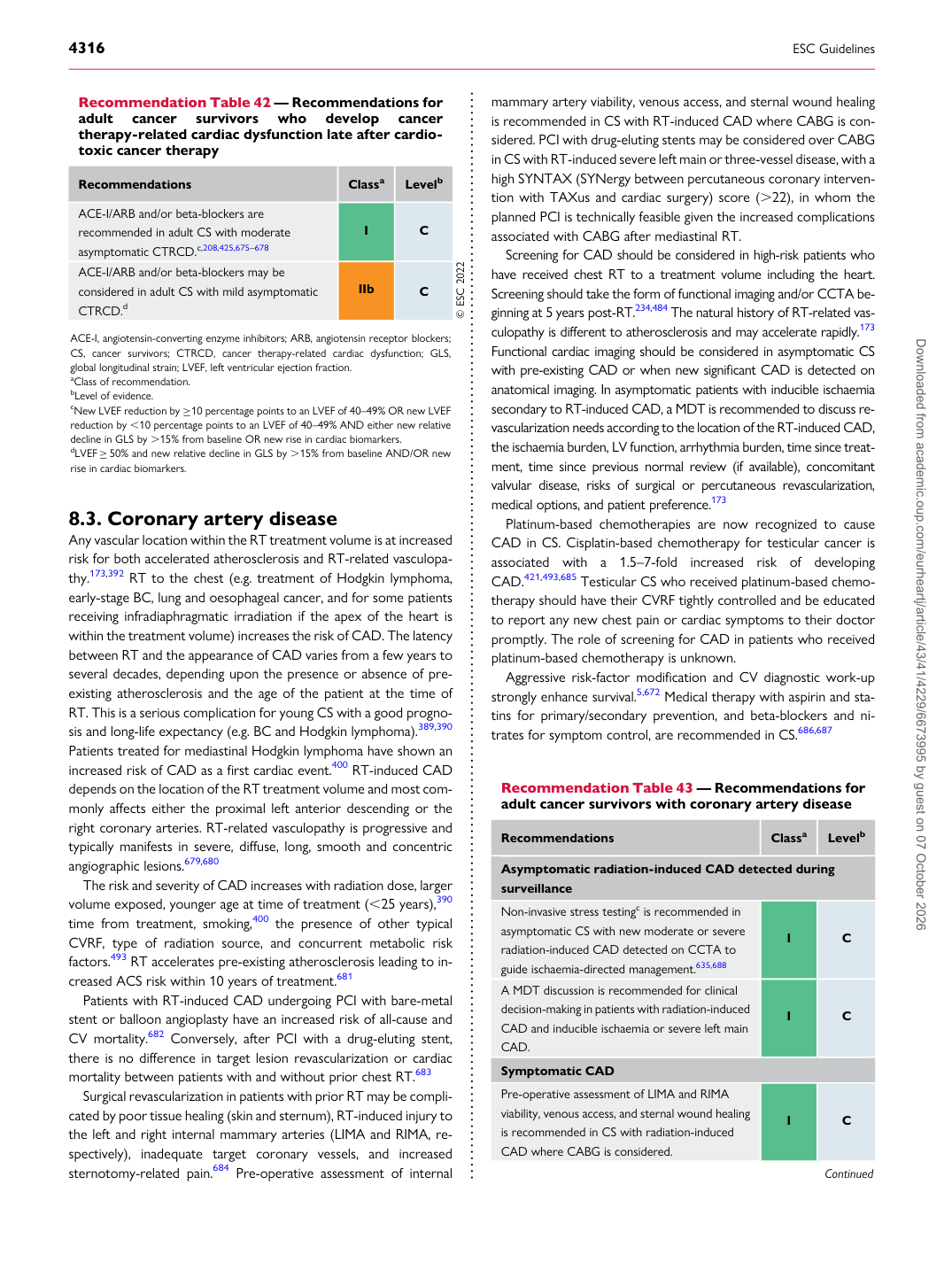
Recommendation Table 42 — Recommendations for adult cancer survivors who develop cancer therapy-related cardiac dysfunction late after cardiotoxic cancer therapy

Recommendations Classa Levelb

ACE-I/ARB and/or beta-blockers are recommended in adult CS with moderate asymptomatic CTRCD.c,208,425,675–678 I C ACE-I/ARB and/or beta-blockers may be considered in adult CS with mild asymptomatic CTRCD.d IIb C © ESC 2022

ACE-I, angiotensin-converting enzyme inhibitors; ARB, angiotensin receptor blockers; CS, cancer survivors; CTRCD, cancer therapy-related cardiac dysfunction; GLS, global longitudinal strain; LVEF, left ventricular ejection fraction. aClass of recommendation. bLevel of evidence. cNew LVEF reduction by ≥10 percentage points to an LVEF of 40–49% OR new LVEF reduction by ,10 percentage points to an LVEF of 40–49% AND either new relative decline in GLS by .15% from baseline OR new rise in cardiac biomarkers. dLVEF ≥50% and new relative decline in GLS by .15% from baseline AND/OR new rise in cardiac biomarkers.

Recommendation Table 43 — Recommendations for adult cancer survivors with coronary artery disease

Recommendations Classa Levelb

Asymptomatic radiation-induced CAD detected during surveillance Non-invasive stress testingc is recommended in asymptomatic CS with new moderate or severe radiation-induced CAD detected on CCTA to guide ischaemia-directed management.635,688 I C A MDT discussion is recommended for clinical decision-making in patients with radiation-induced CAD and inducible ischaemia or severe left main CAD. I C Symptomatic CAD Pre-operative assessment of LIMA and RIMA viability, venous access, and sternal wound healing is recommended in CS with radiation-induced CAD where CABG is considered. I C

Continued Downloaded from academic.oup.com/eurheartj/article/43/41/4229/6673995 by guest on 07 October 2026