p.29 抽出テキスト
5.3 Secondary prevention strategies

Secondary prevention refers to interventions in patients with preexisting CVD, including prior CTR-CVT, and new emerging CTR-CVT during cancer therapy. CVD and comorbidities should receive the optimal therapy before and during cancer therapy as discussed in previous sections. Regular clinical assessments, physical examinations, and CV investigations (including 12-lead ECG, TTE, and cardiac biomarkers) are recommended in patients receiving specific cardiotoxic cancer therapies, with the frequency of surveillance guided by baseline risk and the emergence of new CTR-CVT.5,12,33,53,54,187–190 5.4 Cardiovascular surveillance during

cancer therapies A careful clinical evaluation and physical examination is recommended during cancer treatment to detect early signs and symptoms of CTR-CVT. ECG monitoring is required in patients at risk of cardiac arrhythmias according to specific drug protocols.

5.4.1 Cardiac serum biomarkers

During therapy, NP and cTn should be used for CTRCD screening and diagnosis and they may also serve to guide therapy.55,63,191–194 The release of cTn and NP differ for different cancer treatments. Therefore, an increase in biomarker level should be interpreted in the patient clinical context (cancer treatment timing and comorbidities). It is important to consider that generally accepted cut-offs and reference values of CV biomarkers have not been established for patients with cancer or for those who receive cancer therapies. In addition, levels of NP and cTn may differ according to local laboratories and may be altered by many factors, including age, sex, renal function, obesity, infections, and comorbidities such as AF and pulmonary embolism (PE).53,63,195–197

5.4.2 Cardiac imaging

Cardiac imaging plays a critical role in clinical decision-making during the cancer process.72,198 Imaging techniques—particularly advanced echocardiography and CMR—facilitate early diagnosis and management of CTR-CVT.22,54,94 The frequency of cardiac imaging monitoring during therapy should be adapted according to the estimated baseline risk12 and the expected CTR-CVT manifestation.54 The cardiac imaging technique used should be based on local expertise and availability, and the same imaging modality (i.e. 3D-TTE, 2D-TTE, CMR) is recommended throughout the entire treatment to decrease inter-technique variability.94,199,200 Cardiac imaging should be performed at any time if patients receiving cardiotoxic therapies present with new cardiac symptoms. New definitions of CTRCD are presented in Section 3.1 Early recognition of asymptomatic CTRCD allows clinicians to incorporate cardioprotective therapy before there is a significant decline in LVEF, which may or may not be reversible, and also decreases the risk of interruptions in cancer therapy, which could otherwise affect patients’ survival.22,43,72,94 For the diagnosis and management of asymptomatic CTRCD during cancer treatment, TTE—including 3D-LVEF and GLS assessment—is the preferred technique to detect and confirm cardiac dysfunction.72,83,93,102 GLS evaluation is particularly important in patients with low-normal LVEF to confirm or not asymptomatic myocardial damage.201 It is recommended to use the same vendor to analyse GLS during cancer treatment to accurately compare values over time.73 Therefore, a relative change in GLS has been suggested as the ideal tool to identify asymptomatic mild CTRCD.1,4,94 Different thresholds have been considered in the literature in recent years.93,202,203 Currently, a relative GLS decrease of .15% compared with baseline is the recommended threshold as it reflects the 95% upper

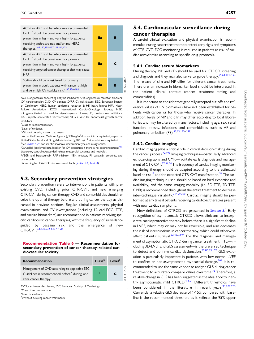
Recommendation Table 6 — Recommendation for secondary prevention of cancer therapy-related cardiovascular toxicity

Recommendation Classa Levelb

Management of CVD according to applicable ESC Guidelines is recommended before,c during, and after cancer therapy. I C © ESC 2022

CVD, cardiovascular disease; ESC, European Society of Cardiology. aClass of recommendation. bLevel of evidence. cWithout delaying cancer treatments.

ACE-I or ARB and beta-blockers recommended for HFf should be considered for primary prevention in high- and very high-risk patients receiving anthracyclines and/or anti-HER2 therapies.145,150,155–157,159,160,175 IIa B ACE-I or ARB and beta-blockers recommended for HFf should be considered for primary prevention in high- and very high-risk patients receiving targeted cancer therapies that may cause HF.g IIa C Statins should be considered for primary prevention in adult patients with cancer at high and very high CV toxicity risk.h,149,176–185 IIa B © ESC 2022

ACE-I, angiotensin-converting enzyme inhibitors; ARB, angiotensin receptor blockers; CV, cardiovascular; CVD, CV disease; CVRF, CV risk factors; ESC, European Society of Cardiology; HER2, human epidermal receptor 2; HF, heart failure; HFA, Heart Failure Association; ICOS, International Cardio-Oncology Society; MEK, mitogen-activated extracellular signal-regulated kinase; PI, proteasome inhibitors; RAF, rapidly accelerated fibrosarcoma; VEGFi, vascular endothelial growth factor inhibitors. aClass of recommendation. bLevel of evidence. cWithout delaying cancer treatments. dAs per the European Medicine Agency: ≥350 mg/m2 doxorubicin or equivalent; as per the United States Food and Drug Administration: ≥300 mg/m2 doxorubicin or equivalent. eSee Section 5.2.1 for specific liposomal doxorubicin type and malignancies. fCarvedilol (preferred beta-blocker for CV protection if there is no contraindication),186 bisoprolol, controlled/extended-release metoprolol succinate and nebivolol. gVEGFi and bevacizumab, RAF inhibitor, MEK inhibitor, PI, dasatinib, ponatinib, and osimertinib. hAccording to HFA-ICOS risk assessment tools (Section 4.1; Table 4). Downloaded from academic.oup.com/eurheartj/article/43/41/4229/6673995 by guest on 07 October 2026