p.25 抽出テキスト
volumes.54,75–79 If 3D echocardiography is not feasible (e.g. unavailable or poor tracking), the modified two-dimensional (2D) Simpson’s biplane method is recommended.80,81 In patients with inadequate TTE image quality, ultrasound-enhancing contrast agents should be added to improve evaluation of LV function and volumes if two or more LV segments are not well visualized.82 Alternatively, in subjects with poor-quality echocardiography windows, when available, CMR should be considered (Figure 8).14,72,83,84 If TTE and CMR are both unavailable for the assessment of LVEF, multigated acquisition nuclear imaging (MUGA) can be considered as a third-line modality. MUGA scans should be avoided whenever possible due to radiation exposure and the inability to obtain other important information (e.g. VHD, PAP, or GLS). Baseline LVEF and GLS are recommended in all patients evaluated with TTE before cardiotoxic cancer treatment initiation to stratify CTR-CVT risk and to identify significant changes during treatment.8,64 Changes in loading conditions occur frequently during chemotherapy (e.g. volume increase due to intravenous [i.v.] fluids, volume loss due to vomiting or diarrhoea, blood pressure [BP] and heart rate changes with pain or stress) and may affect cardiac volumes, LVEF, and GLS quantification. Systemic arterial BP measurement is recommended with all resting TTE as it can influence cardiac function measurements and should be recorded on the TTE report. A baseline borderline (50–54%) or reduced (,50%) LVEF is a risk factor for future CTR-CVT from most cardiotoxic cancer therapies, in particular with anthracyclines or trastuzumab.12,24,74 Increased baseline indexed LV end-diastolic volume can be a predictor of major CV events (symptomatic HF or cardiac death) during anthracycline chemotherapy in patients with preserved LVEF.85 A normal LVEF does not exclude CTRCD and deformation parameters can detect early systolic impairment with sufficient test reliability.86–89 Determination of GLS using speckle tracking is recommended at baseline, using three apical views,90 particularly in moderate- and high-risk patients. Baseline GLS can predict LVD89–94 in patients receiving anthracyclines and/or trastuzumab. Strain measurements may be subject to inter-vendor variability95 and serial GLS measurement for each patient is recommended to be performed using the same machine/software. A median GLS change of 13.6% predicted a future fall in LVEF with a 95% upper limit of GLS reduction of 15%.93 Using the 15% cut-off improves specificity and is therefore the threshold recommended when monitoring GLS during cancer therapy. Global circumferential strain96 has been reported to identify patients at risk of CTRCD, but data are currently insufficient to recommend its use routinely. Baseline LV diastolic function may be associated with a small risk of subsequent systolic dysfunction, especially with anthracyclines and trastuzumab, although the evidence is not consistent.97,98 Chest computed tomography (CT) or CMR may be helpful for identifying subclinical CVD such as coronary calcium or intracardiac masses on readily available routine imaging performed for cancer staging.99 In the secondary prevention setting or patients with symptoms or signs of pre-existing CVD, a careful evaluation should begin with a comprehensive TTE.73 This is both to obtain baseline assessment as in the primary prevention setting and to determine the severity of the underlying CVD. In case of poor-quality or uninterpretable TTE images, or if a specific CVD is identified (e.g. hypertrophic cardiomyopathy), CMR should be considered for further risk assessment.

Functional imaging tests for myocardial ischaemia—including stress echocardiography, perfusion CMR, or nuclear myocardial perfusion imaging—should be performed to assess for ischaemia in symptomatic patients (stable angina, limiting dyspnoea) if clinical suspicion of coronary artery disease (CAD) exists, especially prior to use of cancer therapies associated with vascular toxicity (e.g. fluoropyrimidines, VEGFi, breakpoint cluster region–Abelson oncogene locus [BCR-ABL], tyrosine kinase inhibitors [TKI]). Alternatively, in patients with low to intermediate pre-test probability of CAD, CCTA is a robust alternate modality with high sensitivity to rule out obstructive CAD.100,101

4.6 Cardiopulmonary fitness assessment

Maximal cardiopulmonary exercise testing (CPET) assesses the integrative capacity of the CV system to transport oxygen and energy substrate to skeletal muscle during exercise,109 described as cardiorespiratory fitness (CRF). CPET can therefore provide a more global assessment of CV health than organ-specific tools. CPET-derived

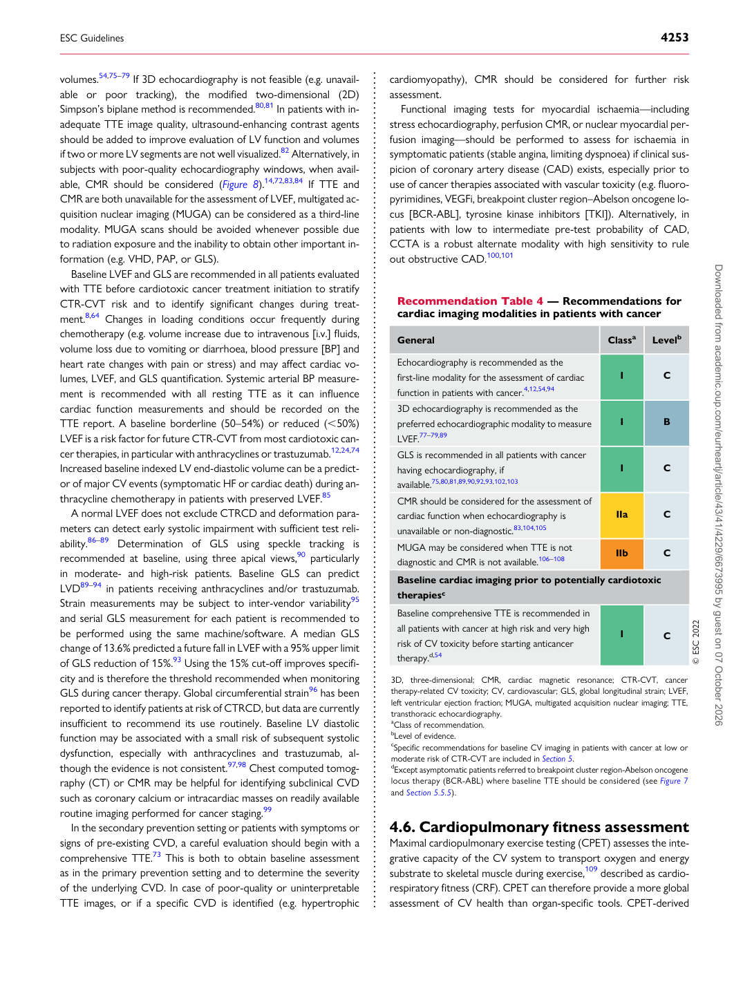
Recommendation Table 4 — Recommendations for cardiac imaging modalities in patients with cancer

General Classa Levelb

Echocardiography is recommended as the first-line modality for the assessment of cardiac function in patients with cancer.4,12,54,94 I C 3D echocardiography is recommended as the preferred echocardiographic modality to measure LVEF.77–79,89 I B GLS is recommended in all patients with cancer having echocardiography, if available.75,80,81,89,90,92,93,102,103 I C CMR should be considered for the assessment of cardiac function when echocardiography is unavailable or non-diagnostic.83,104,105 IIa C MUGA may be considered when TTE is not diagnostic and CMR is not available.106–108 IIb C Baseline cardiac imaging prior to potentially cardiotoxic therapiesc Baseline comprehensive TTE is recommended in all patients with cancer at high risk and very high risk of CV toxicity before starting anticancer therapy.d,54 I C © ESC 2022

3D, three-dimensional; CMR, cardiac magnetic resonance; CTR-CVT, cancer therapy-related CV toxicity; CV, cardiovascular; GLS, global longitudinal strain; LVEF, left ventricular ejection fraction; MUGA, multigated acquisition nuclear imaging; TTE, transthoracic echocardiography. aClass of recommendation. bLevel of evidence. cSpecific recommendations for baseline CV imaging in patients with cancer at low or moderate risk of CTR-CVT are included in Section 5. dExcept asymptomatic patients referred to breakpoint cluster region-Abelson oncogene locus therapy (BCR-ABL) where baseline TTE should be considered (see Figure 7 and Section 5.5.5). Downloaded from academic.oup.com/eurheartj/article/43/41/4229/6673995 by guest on 07 October 2026