p.24 抽出テキスト
4.5 Cardiovascular imaging

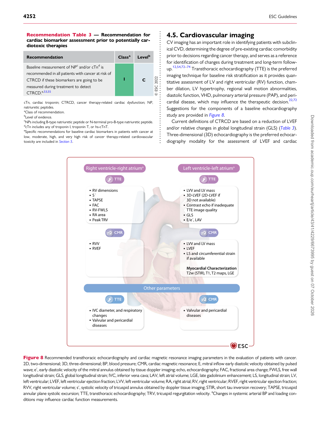
CV imaging has an important role in identifying patients with subclinical CVD, determining the degree of pre-existing cardiac comorbidity prior to decisions regarding cancer therapy, and serves as a reference for identification of changes during treatment and long-term followup.12,54,72–74 Transthoracic echocardiography (TTE) is the preferred imaging technique for baseline risk stratification as it provides quantitative assessment of LV and right ventricular (RV) function, chamber dilation, LV hypertrophy, regional wall motion abnormalities, diastolic function, VHD, pulmonary arterial pressure (PAP), and pericardial disease, which may influence the therapeutic decision.22,72 Suggestions for the components of a baseline echocardiography study are provided in Figure 8. Current definitions of CTRCD are based on a reduction of LVEF and/or relative changes in global longitudinal strain (GLS) (Table 3). Three-dimensional (3D) echocardiography is the preferred echocardiography modality for the assessment of LVEF and cardiac

Recommendation Table 3 — Recommendation for cardiac biomarker assessment prior to potentially cardiotoxic therapies

Recommendation Classa Levelb Baseline measurement of NPc and/or cTnd is recommended in all patients with cancer at risk of CTRCD if these biomarkers are going to be measured during treatment to detect CTRCD.e,53,55 I C © ESC 2022

cTn, cardiac troponin; CTRCD, cancer therapy-related cardiac dysfunction; NP, natriuretic peptides. aClass of recommendation. bLevel of evidence. cNPs including B-type natriuretic peptide or N-terminal pro-B-type natriuretic peptide. dcTn includes any of troponin I, troponin T, or hs-cTnT. eSpecific recommendations for baseline cardiac biomarkers in patients with cancer at low, moderate, high, and very high risk of cancer therapy-related cardiovascular toxicity are included in Section 5.

TTE Left ventricle-left atriuma

TTE

LVV and LV mass 3D-LVEF (2D-LVEF if 3D not available) Contrast echo if inadequate TTE image quality GLS E/e ́, LAV

RVV RVEF CMR

Right ventricle-right atriuma

RV dimensions S ́ TAPSE FAC RV-FWLS RA area Peak TRV

LVV and LV mass LVEF LS and circumferential strain if available

Myocardial Characterization T2w (STIR), T1, T2 maps, LGE

CMR

Valvular and pericardial diseases

IVC diameter, and respiratory changes Valvular and pericardial diseases Other parameters

TTE CMR

Figure 8 Recommended transthoracic echocardiography and cardiac magnetic resonance imaging parameters in the evaluation of patients with cancer. 2D, two-dimensional; 3D, three-dimensional; BP, blood pressure; CMR, cardiac magnetic resonance; E, mitral inflow early diastolic velocity obtained by pulsed wave; e′, early diastolic velocity of the mitral annulus obtained by tissue doppler imaging; echo, echocardiography; FAC, fractional area change; FWLS, free wall longitudinal strain; GLS, global longitudinal strain; IVC, inferior vena cava; LAV, left atrial volume; LGE, late gadolinium enhancement; LS, longitudinal strain; LV, left ventricular; LVEF, left ventricular ejection fraction; LVV, left ventricular volume; RA, right atrial; RV, right ventricular; RVEF, right ventricular ejection fraction; RVV, right ventricular volume; s′, systolic velocity of tricuspid annulus obtained by doppler tissue imaging; STIR, short tau inversion recovery; TAPSE, tricuspid annular plane systolic excursion; TTE, transthoracic echocardiography; TRV, tricuspid regurgitation velocity. aChanges in systemic arterial BP and loading conditions may influence cardiac function measurements. Downloaded from academic.oup.com/eurheartj/article/43/41/4229/6673995 by guest on 07 October 2026