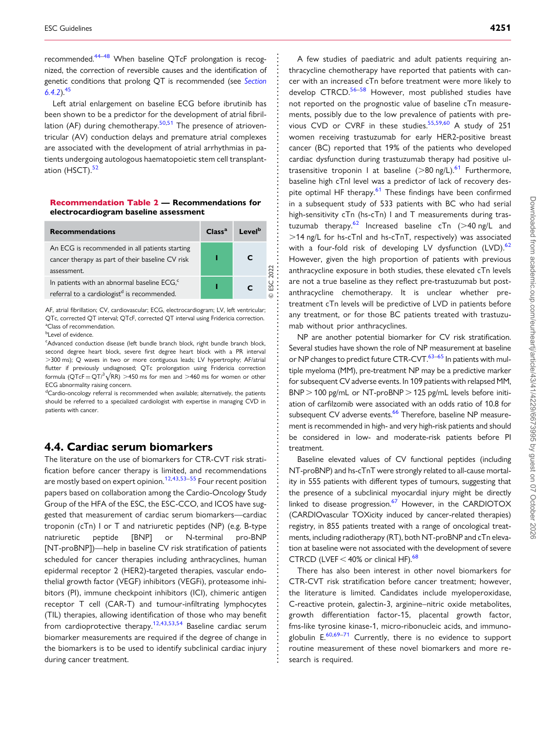
p.23 抽出テキスト
recommended.44–48 When baseline QTcF prolongation is recognized, the correction of reversible causes and the identification of genetic conditions that prolong QT is recommended (see Section 6.4.2).45 Left atrial enlargement on baseline ECG before ibrutinib has been shown to be a predictor for the development of atrial fibrillation (AF) during chemotherapy.50,51 The presence of atrioventricular (AV) conduction delays and premature atrial complexes are associated with the development of atrial arrhythmias in patients undergoing autologous haematopoietic stem cell transplantation (HSCT).52

4.4 Cardiac serum biomarkers

The literature on the use of biomarkers for CTR-CVT risk stratification before cancer therapy is limited, and recommendations are mostly based on expert opinion.12,43,53–55 Four recent position papers based on collaboration among the Cardio-Oncology Study Group of the HFA of the ESC, the ESC-CCO, and ICOS have suggested that measurement of cardiac serum biomarkers—cardiac troponin (cTn) I or T and natriuretic peptides (NP) (e.g. B-type natriuretic peptide [BNP] or N-terminal pro-BNP [NT-proBNP])—help in baseline CV risk stratification of patients scheduled for cancer therapies including anthracyclines, human epidermal receptor 2 (HER2)-targeted therapies, vascular endothelial growth factor (VEGF) inhibitors (VEGFi), proteasome inhibitors (PI), immune checkpoint inhibitors (ICI), chimeric antigen receptor T cell (CAR-T) and tumour-infiltrating lymphocytes (TIL) therapies, allowing identification of those who may benefit from cardioprotective therapy.12,43,53,54 Baseline cardiac serum biomarker measurements are required if the degree of change in the biomarkers is to be used to identify subclinical cardiac injury during cancer treatment. A few studies of paediatric and adult patients requiring anthracycline chemotherapy have reported that patients with cancer with an increased cTn before treatment were more likely to develop CTRCD.56–58 However, most published studies have not reported on the prognostic value of baseline cTn measurements, possibly due to the low prevalence of patients with previous CVD or CVRF in these studies.55,59,60 A study of 251 women receiving trastuzumab for early HER2-positive breast cancer (BC) reported that 19% of the patients who developed cardiac dysfunction during trastuzumab therapy had positive ultrasensitive troponin I at baseline (.80 ng/L).61 Furthermore, baseline high cTnI level was a predictor of lack of recovery despite optimal HF therapy.61 These findings have been confirmed in a subsequent study of 533 patients with BC who had serial high-sensitivity cTn (hs-cTn) I and T measurements during trastuzumab therapy.62 Increased baseline cTn (.40 ng/L and .14 ng/L for hs-cTnI and hs-cTnT, respectively) was associated with a four-fold risk of developing LV dysfunction (LVD).62

However, given the high proportion of patients with previous anthracycline exposure in both studies, these elevated cTn levels are not a true baseline as they reflect pre-trastuzumab but postanthracycline chemotherapy. It is unclear whether pretreatment cTn levels will be predictive of LVD in patients before any treatment, or for those BC patients treated with trastuzumab without prior anthracyclines. NP are another potential biomarker for CV risk stratification. Several studies have shown the role of NP measurement at baseline or NP changes to predict future CTR-CVT.63–65 In patients with multiple myeloma (MM), pre-treatment NP may be a predictive marker for subsequent CV adverse events. In 109 patients with relapsed MM, BNP . 100 pg/mL or NT-proBNP . 125 pg/mL levels before initiation of carfilzomib were associated with an odds ratio of 10.8 for subsequent CV adverse events.66 Therefore, baseline NP measurement is recommended in high- and very high-risk patients and should be considered in low- and moderate-risk patients before PI treatment. Baseline elevated values of CV functional peptides (including NT-proBNP) and hs-cTnT were strongly related to all-cause mortality in 555 patients with different types of tumours, suggesting that the presence of a subclinical myocardial injury might be directly linked to disease progression.67 However, in the CARDIOTOX (CARDIOvascular TOXicity induced by cancer-related therapies) registry, in 855 patients treated with a range of oncological treatments, including radiotherapy (RT), both NT-proBNP and cTn elevation at baseline were not associated with the development of severe CTRCD (LVEF , 40% or clinical HF).68 There has also been interest in other novel biomarkers for CTR-CVT risk stratification before cancer treatment; however, the literature is limited. Candidates include myeloperoxidase, C-reactive protein, galectin-3, arginine–nitric oxide metabolites, growth differentiation factor-15, placental growth factor, fms-like tyrosine kinase-1, micro-ribonucleic acids, and immunoglobulin E.60,69–71 Currently, there is no evidence to support routine measurement of these novel biomarkers and more research is required.

Recommendation Table 2 — Recommendations for electrocardiogram baseline assessment

Recommendations Classa Levelb

An ECG is recommended in all patients starting cancer therapy as part of their baseline CV risk assessment. I C In patients with an abnormal baseline ECG,c referral to a cardiologistd is recommended. I C © ESC 2022

AF, atrial fibrillation; CV, cardiovascular; ECG, electrocardiogram; LV, left ventricular; QTc, corrected QT interval; QTcF, corrected QT interval using Fridericia correction. aClass of recommendation. bLevel of evidence. cAdvanced conduction disease (left bundle branch block, right bundle branch block, second degree heart block, severe first degree heart block with a PR interval .300 ms); Q waves in two or more contiguous leads; LV hypertrophy; AF/atrial flutter if previously undiagnosed; QTc prolongation using Fridericia correction formula (QTcF = QT/3√RR) .450 ms for men and .460 ms for women or other ECG abnormality raising concern. dCardio-oncology referral is recommended when available; alternatively, the patients should be referred to a specialized cardiologist with expertise in managing CVD in patients with cancer. Downloaded from academic.oup.com/eurheartj/article/43/41/4229/6673995 by guest on 07 October 2026