p.21 抽出テキスト
4.2 History and clinical examination

A careful clinical history and physical examination is recommended as part of the baseline risk assessment. Oncology patients can be divided into two cohorts with respect to the presence or absence of pre-existing CVD. A primary prevention strategy can be considered in patients without previous CVD or CTR-CVT while secondary prevention includes interventions in patients with prior or active CVD or previous CTR-CVT.12

Reviewing traditional risk factors for CVD is recommended. Where present, the efficacy of treatment and control of these modifiable risk factors should be determined to ensure optimal control during cancer therapy.4,34 Although recent SCORE2 and SCORE2-OP19 tables are not focused on patients with cancer, risk calculation is recommended for patients with cancer .40 years of age (unless they are automatically categorized as being at high risk or very high risk based on documented CVD, diabetes mellitus [DM], kidney disease, or a highly elevated single risk factor) as a reference to optimize CVRF treatment goals.19,31,35 A family history of premature CVD should be considered because genetic abnormalities associated with CVD may predispose patients with cancer to a higher risk of CTR-CVT.36–38 Lifestyle factors such as smoking, alcohol consumption, sedentary lifestyle, exposure to pollution, and frailty are important shared risk factors for both cancer and CVD. Information on prior history of cancer, cardiotoxic cancer therapies, and their respective doses should be collected. Patients should be asked about typical cardiac symptoms (e.g. chest pain with activity, dyspnoea on exertion, orthopnoea, palpitations, and peripheral oedema), which can guide clinical examination and investigations. Physical examination should document vital signs and look for potential indicators of undiagnosed CVD such as HF, pericardial disease, VHD, and arrhythmias.39–42 The second scenario is secondary prevention in patients with a prior history of CVD. These patients with cancer are potentially at high or very high risk of future CV events,12 and require a more comprehensive clinical evaluation of their CVD, its severity, and prior and current treatments. Depending on the type and severity of CVD, additional investigations—including resting or stress echocardiography, cardiac magnetic resonance (CMR), nuclear perfusion imaging, and coronary computed tomography angiography (CCTA) —may be indicated to determine risk status. Identifying prior CVD should not automatically be a reason to withhold cancer therapy but considered an opportunity to optimize CV risk prior to and during treatment. Risk/benefit discussions should include the patient, oncologist or haematologist, and—where available—a specialized cardio-oncology service. Additional factors that add to the complexity of baseline CV risk assessment are the cancer type and prognosis, and type, duration, and intensity of cancer treatment (Figure 1).4,12,43 Clinical history, physical examination features, and treatment-related risk factors that contribute to CTR-CVT for various cancer therapies are summarized in Supplementary data, Table S8. These risk factors should be collected and considered along with baseline ECG, cardiac serum biomarkers, and cardiac imaging tests (summarized in Figure 7) to complete baseline CTR-CVT evaluation.

4.3 Electrocardiogram

A baseline 12-lead ECG is a readily available test that can provide important clues to underlying CVD. ECG evidence of chamber enlargement, conduction abnormalities, arrhythmias, ischaemia, or prior myocardial infarction (MI), and low voltages should be interpreted in the clinical context. A baseline ECG is recommended prior to starting a cancer treatment known to cause QTc prolongation.44–49 Measurement of QTc using the Fridericia correction (QTcF) is

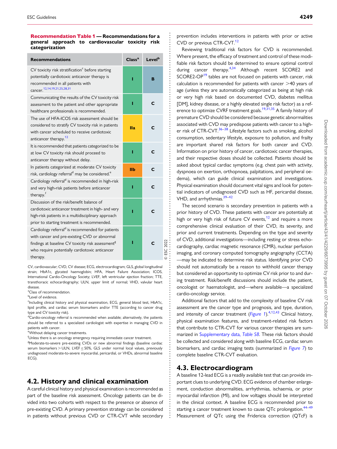
Recommendation Table 1 — Recommendations for a general approach to cardiovascular toxicity risk categorization

Recommendations Classa Levelb CV toxicity risk stratificationc before starting potentially cardiotoxic anticancer therapy is recommended in all patients with cancer.12,14,19,21,25,28,31 I B Communicating the results of the CV toxicity risk assessment to the patient and other appropriate healthcare professionals is recommended. I C The use of HFA-ICOS risk assessment should be considered to stratify CV toxicity risk in patients with cancer scheduled to receive cardiotoxic anticancer therapy.12 IIa C It is recommended that patients categorized to be at low CV toxicity risk should proceed to anticancer therapy without delay. I C In patients categorized at moderate CV toxicity risk, cardiology referrald may be considered.e IIb C Cardiology referrald is recommended in high-risk and very high-risk patients before anticancer therapy.f I C Discussion of the risk/benefit balance of cardiotoxic anticancer treatment in high- and very high-risk patients in a multidisciplinary approach prior to starting treatment is recommended. I C Cardiology referrald is recommended for patients with cancer and pre-existing CVD or abnormal findings at baseline CV toxicity risk assessmentg who require potentially cardiotoxic anticancer therapy. I C © ESC 2022

CV, cardiovascular; CVD, CV disease; ECG, electrocardiogram; GLS, global longitudinal strain; HbA1c, glycated haemoglobin; HFA, Heart Failure Association; ICOS, International Cardio-Oncology Society; LVEF, left ventricular ejection fraction; TTE, transthoracic echocardiography; ULN, upper limit of normal; VHD, valvular heart disease. aClass of recommendation. bLevel of evidence. cIncluding clinical history and physical examination, ECG, general blood test, HbA1c, lipid profile, and cardiac serum biomarkers and/or TTE (according to cancer drug type and CV toxicity risk). dCardio-oncology referral is recommended when available; alternatively, the patients should be referred to a specialized cardiologist with expertise in managing CVD in patients with cancer. eWithout delaying cancer treatments. fUnless there is an oncology emergency requiring immediate cancer treatment. gModerate-to-severe pre-existing CVDs or new abnormal findings (baseline cardiac serum biomarkers . ULN, LVEF ≤50%, GLS under normal local values, previously undiagnosed moderate-to-severe myocardial, pericardial, or VHDs, abnormal baseline ECG). Downloaded from academic.oup.com/eurheartj/article/43/41/4229/6673995 by guest on 07 October 2026