p.17 抽出テキスト
Only a limited number of retrospective risk scores have been published in patients with cancer. Most of these scores have been developed for specific cancer-patient groups and cannot be readily applied or extrapolated to other type of malignancies.24–29 While further validation is needed, HFA-ICOS risk assessment tools should be considered to determine pre-treatment risk of CTR-CVT as they are easy to use and implement in oncology and haematology services (Table 4; Supplementary data, Tables S2–S7).12,13 Other CV risk

Physical and metabolic

assessmentb ECG (Class I) Pre-existing CVD or

at risk of CTRCDc

New, untreated or uncontrolled medical, and

lifestyle CVRF

Arrhythmias Prolonged QTcF

Check and correct precipitant factorsf

(Class I) Ref cardiologistg

(Class I) Ref rdiologist g

(Class I)

Suspected

CVD cTn and NP

increasedd TTE (LVEF and GLS)

reducede

Aggressive management of CVRF and pre-existing CVD according to ESC Guidelines Healthy lifestyle promotion

(Class I)

Baseline cardiovascular toxicity risk assessment

Consider primary vs secondary preventive strategies according to CV toxicity risk,

pre-existing CVD, and cancer treatment type

HFA-ICOS risk assessment: CVRFa CVD history Cancer history Cancer treatment history

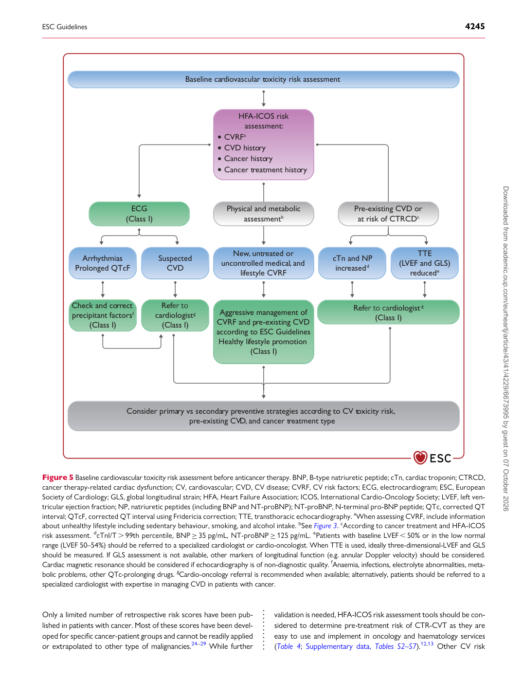
Figure 5 Baseline cardiovascular toxicity risk assessment before anticancer therapy. BNP, B-type natriuretic peptide; cTn, cardiac troponin; CTRCD, cancer therapy-related cardiac dysfunction; CV, cardiovascular; CVD, CV disease; CVRF, CV risk factors; ECG, electrocardiogram; ESC, European Society of Cardiology; GLS, global longitudinal strain; HFA, Heart Failure Association; ICOS, International Cardio-Oncology Society; LVEF, left ventricular ejection fraction; NP, natriuretic peptides (including BNP and NT-proBNP); NT-proBNP, N-terminal pro-BNP peptide; QTc, corrected QT interval; QTcF, corrected QT interval using Fridericia correction; TTE, transthoracic echocardiography. aWhen assessing CVRF, include information about unhealthy lifestyle including sedentary behaviour, smoking, and alcohol intake. bSee Figure 3. cAccording to cancer treatment and HFA-ICOS risk assessment. dcTnI/T . 99th percentile, BNP ≥35 pg/mL, NT-proBNP ≥125 pg/mL. ePatients with baseline LVEF , 50% or in the low normal range (LVEF 50–54%) should be referred to a specialized cardiologist or cardio-oncologist. When TTE is used, ideally three-dimensional-LVEF and GLS should be measured. If GLS assessment is not available, other markers of longitudinal function (e.g. annular Doppler velocity) should be considered. Cardiac magnetic resonance should be considered if echocardiography is of non-diagnostic quality. fAnaemia, infections, electrolyte abnormalities, metabolic problems, other QTc-prolonging drugs. gCardio-oncology referral is recommended when available; alternatively, patients should be referred to a specialized cardiologist with expertise in managing CVD in patients with cancer. Downloaded from academic.oup.com/eurheartj/article/43/41/4229/6673995 by guest on 07 October 2026