p.16 抽出テキスト
4 Cardiovascular toxicity risk

stratification before anticancer therapy

The optimal time to consider CVD prevention strategies in patients with cancer is at the time of cancer diagnosis and prior to the initiation of cancer treatment.4,5 This enables the oncology team to consider CV risk while making cancer treatment choices, educating patients regarding their CV risk, personalizing CV surveillance and follow-up strategies, and making appropriate referrals of high-risk patients to cardio-oncology services. These strategies are needed to mitigate CVD risk, and improve the adherence to effective cancer treatments and the overall survival. CVD prevention strategies require a personalized approach. Risk assessment is a challenging task and it is vital that clinicians adopt a systematic approach without delaying oncological treatment.12,21,22

Figure 5 provides a comprehensive approach to risk assessment. The choice of the cardiac tests (electrocardiogram [ECG], biomarkers, and imaging) should be individualized based on CV risk and the planned cancer treatments.

4.1 General approach to cardiovascular

toxicity risk in patients with cancer Pre-treatment CTR-CVT risk assessment should ideally be performed using a recognized risk stratification method where multiple risk factors are incorporated to determine patient-specific risk.23

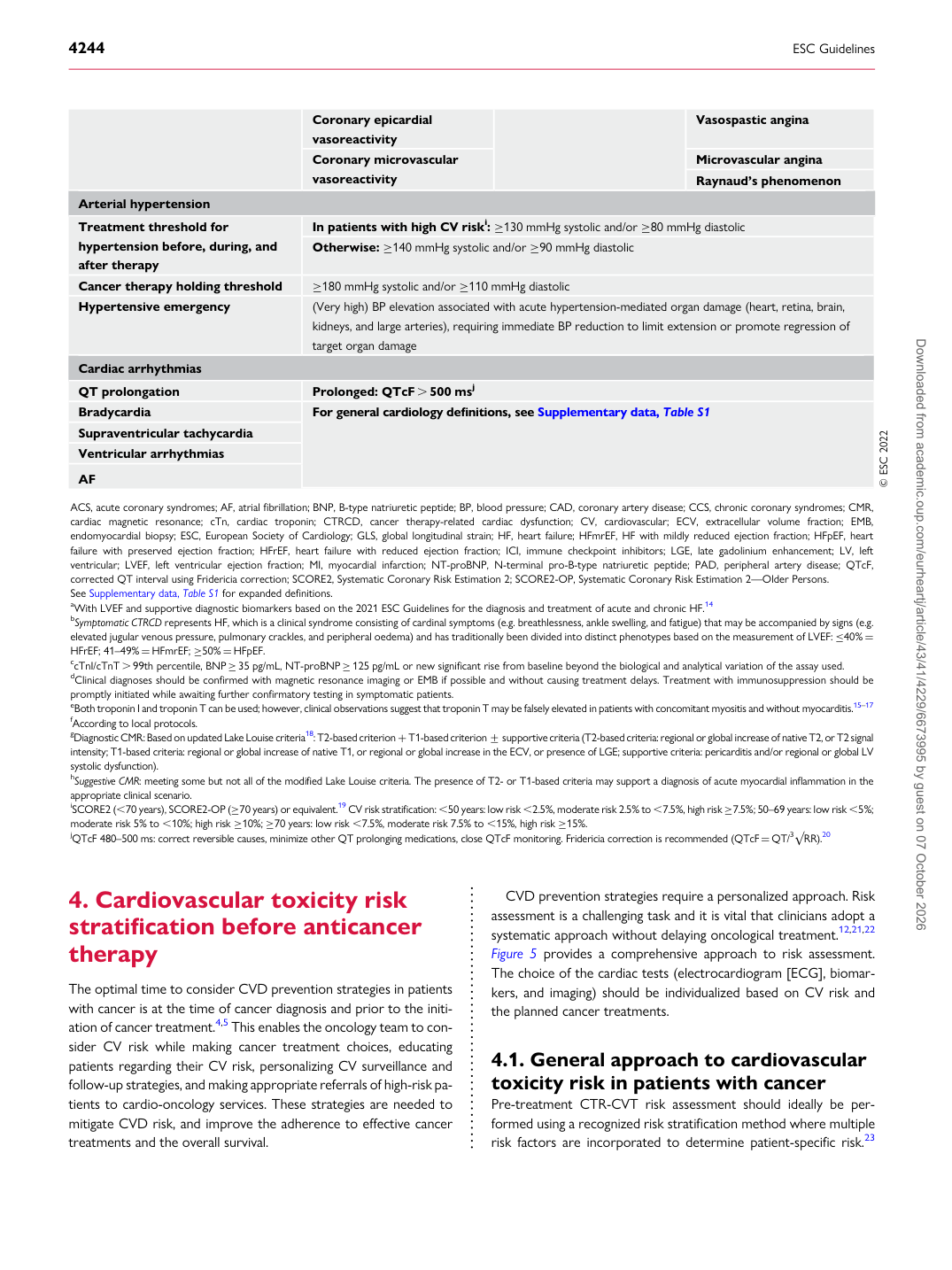
Coronary epicardial vasoreactivity Vasospastic angina Coronary microvascular vasoreactivity Microvascular angina

Raynaud’s phenomenon

Arterial hypertension Treatment threshold for hypertension before, during, and after therapy In patients with high CV riski: ≥130 mmHg systolic and/or ≥80 mmHg diastolic

Otherwise: ≥140 mmHg systolic and/or ≥90 mmHg diastolic

Cancer therapy holding threshold ≥180 mmHg systolic and/or ≥110 mmHg diastolic Hypertensive emergency (Very high) BP elevation associated with acute hypertension-mediated organ damage (heart, retina, brain, kidneys, and large arteries), requiring immediate BP reduction to limit extension or promote regression of

target organ damage

Cardiac arrhythmias QT prolongation Prolonged: QTcF . 500 msj Bradycardia For general cardiology definitions, see Supplementary data, Table S1 Supraventricular tachycardia Ventricular arrhythmias AF © ESC 2022

ACS, acute coronary syndromes; AF, atrial fibrillation; BNP, B-type natriuretic peptide; BP, blood pressure; CAD, coronary artery disease; CCS, chronic coronary syndromes; CMR, cardiac magnetic resonance; cTn, cardiac troponin; CTRCD, cancer therapy-related cardiac dysfunction; CV, cardiovascular; ECV, extracellular volume fraction; EMB, endomyocardial biopsy; ESC, European Society of Cardiology; GLS, global longitudinal strain; HF, heart failure; HFmrEF, HF with mildly reduced ejection fraction; HFpEF, heart failure with preserved ejection fraction; HFrEF, heart failure with reduced ejection fraction; ICI, immune checkpoint inhibitors; LGE, late gadolinium enhancement; LV, left ventricular; LVEF, left ventricular ejection fraction; MI, myocardial infarction; NT-proBNP, N-terminal pro-B-type natriuretic peptide; PAD, peripheral artery disease; QTcF, corrected QT interval using Fridericia correction; SCORE2, Systematic Coronary Risk Estimation 2; SCORE2-OP, Systematic Coronary Risk Estimation 2—Older Persons. See Supplementary data, Table S1 for expanded definitions. aWith LVEF and supportive diagnostic biomarkers based on the 2021 ESC Guidelines for the diagnosis and treatment of acute and chronic HF.14 bSymptomatic CTRCD represents HF, which is a clinical syndrome consisting of cardinal symptoms (e.g. breathlessness, ankle swelling, and fatigue) that may be accompanied by signs (e.g. elevated jugular venous pressure, pulmonary crackles, and peripheral oedema) and has traditionally been divided into distinct phenotypes based on the measurement of LVEF: ≤40% = HFrEF; 41–49% = HFmrEF; ≥50% = HFpEF. ccTnI/cTnT . 99th percentile, BNP ≥35 pg/mL, NT-proBNP ≥125 pg/mL or new significant rise from baseline beyond the biological and analytical variation of the assay used. dClinical diagnoses should be confirmed with magnetic resonance imaging or EMB if possible and without causing treatment delays. Treatment with immunosuppression should be promptly initiated while awaiting further confirmatory testing in symptomatic patients. eBoth troponin I and troponin T can be used; however, clinical observations suggest that troponin T may be falsely elevated in patients with concomitant myositis and without myocarditis.15–17 fAccording to local protocols. gDiagnostic CMR: Based on updated Lake Louise criteria18: T2-based criterion + T1-based criterion + supportive criteria (T2-based criteria: regional or global increase of native T2, or T2signal intensity; T1-based criteria: regional or global increase of native T1, or regional or global increase in the ECV, or presence of LGE; supportive criteria: pericarditis and/or regional or global LV systolic dysfunction). hSuggestive CMR: meeting some but not all of the modified Lake Louise criteria. The presence of T2- or T1-based criteria may support a diagnosis of acute myocardial inflammation in the appropriate clinical scenario. iSCORE2 (,70 years), SCORE2-OP (≥70 years) or equivalent.19 CV risk stratification: ,50 years: low risk ,2.5%, moderate risk 2.5% to ,7.5%, high risk ≥7.5%; 50–69 years: low risk ,5%; moderate risk 5% to ,10%; high risk ≥10%; ≥70 years: low risk ,7.5%, moderate risk 7.5% to ,15%, high risk ≥15%. jQTcF 480–500 ms: correct reversible causes, minimize other QT prolonging medications, close QTcF monitoring. Fridericia correction is recommended (QTcF= QT/3√RR).20 Downloaded from academic.oup.com/eurheartj/article/43/41/4229/6673995 by guest on 07 October 2026