p.45 抽出テキスト
Suspicion of thrombosis

Management of left-sided obstructive and non-obstructive mechanical prosthetic thrombosis

Y Echo (TTE + TOE/fluoroscopy)

Obstructive thrombosisa

Disappearance or decrease of thrombus

Recent inadequate

anticoagulation Surgery immediately

available

High-risk for surgery

Surgery Fibrinolysis Follow-up

i.v. UFH ± ASA Surgery Fibrinolysis

Success/failure Non-obstructive thrombosis

Large thrombus

(≥ 10 mm) Large thrombus

(≥ 10 mm)

Persistence of thrombus or TE

Surgery (or fibrinolysis if surgery is high risk)

Optimize anticoagulation (follow-up)

Signs of TE

Critically ill

Thromboembolism (clinical/cerebral imaging)

FAILURE

N

N Y SUCCESS

Y N Y N

Y N

Optimize anticoagulation

Y N

Follow-up

N Y

Optimize anticoagulation (follow-up)

Y N

Persistence of thrombus

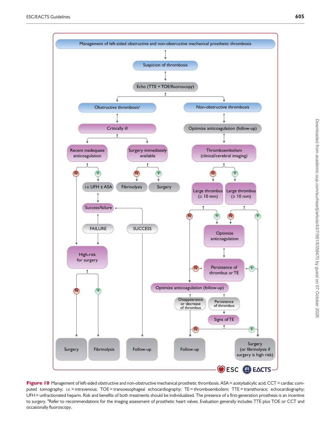
Figure 10 Management of left-sided obstructive and non-obstructive mechanical prosthetic thrombosis. ASA = acetylsalicylic acid; CCT = cardiac computed tomography; i.v.= intravenous; TOE= transoesophageal echocardiography; TE = thromboembolism; TTE = transthoracic echocardiography; UFH= unfractionated heparin. Risk and benefits of both treatments should be individualized. The presence of a first-generation prosthesis is an incentive to surgery. aRefer to recommendations for the imaging assessment of prosthetic heart valves. Evaluation generally includes TTE plus TOE or CCT and occasionally fluoroscopy. Downloaded from academic.oup.com/eurheartj/article/43/7/561/6358470 by guest on 07 October 2026