p.22 抽出テキスト
Valve morphology by echocardiography suspicious of AS

AVA ≤ 1.0 cm2

Reassess under normal flow conditions

N

N

Y

CCT to assess AV calcificatione Pseudo-severe ASd Severe AS Severe AS

Assess velocity/gradient

Low-gradient AS Vmax < 4 m/s, ΔPm < 40 mmHg High-gradient AS

Vmax ≥ 4 m/s, ΔPm ≥ 40 mmHg

High flow statusa Moderate AS

N Y

High flow status

reversible Check blood pressure and exclude measurement errors that may cause underestimation of gradient, flow or AVA

Y Define flow statusa

Low flow SVi ≤ 35 mL/m2 Normal flow SVi > 35 mL/m2

Severe AS unlikely

LVEF ≥ 50%

Y

DSE flow reservec

N

AVA ≤ 1.0 cm2

Y Y Integrated approachb

N

N

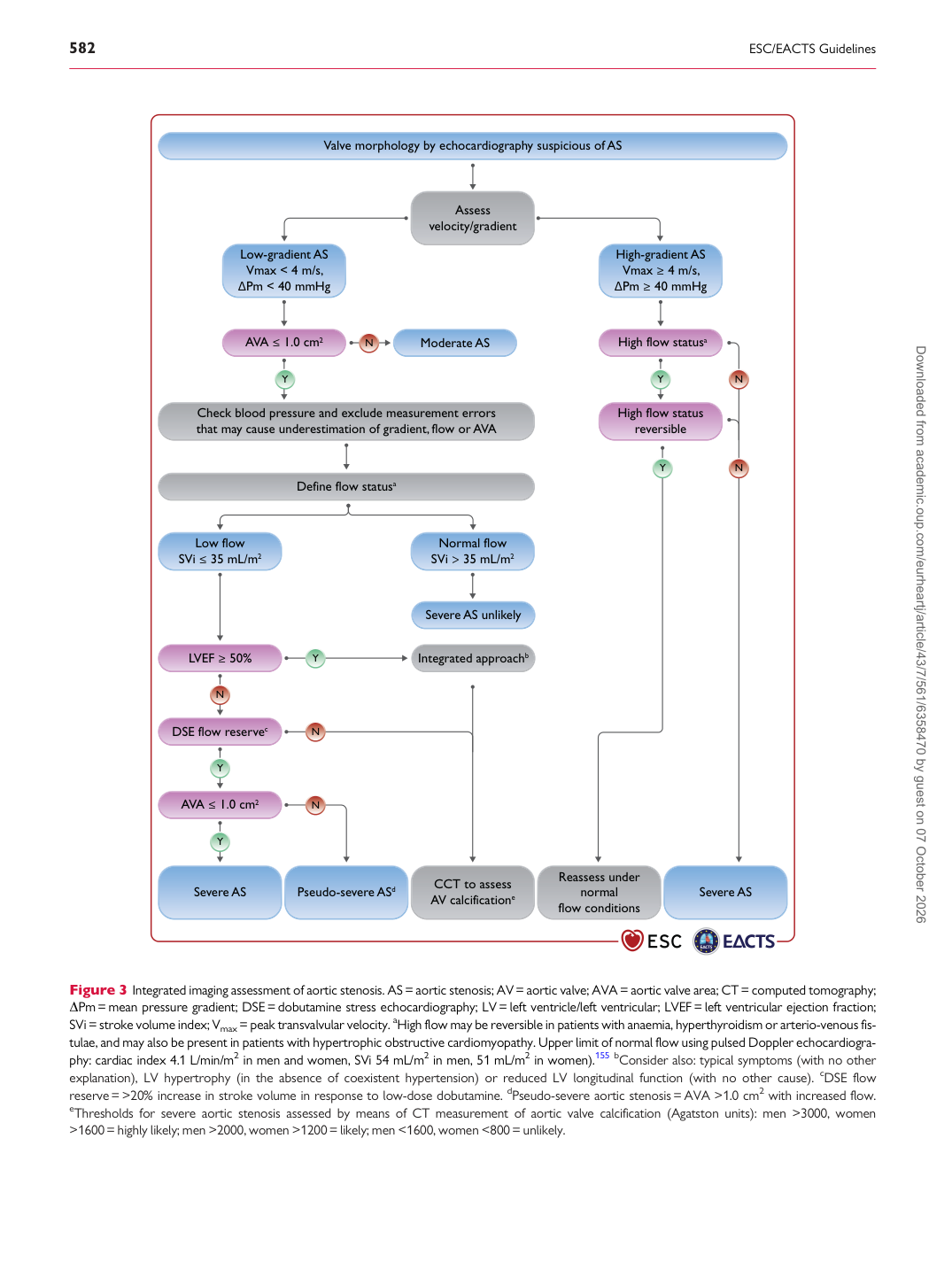
Figure 3 Integrated imaging assessment of aortic stenosis. AS= aortic stenosis; AV= aortic valve; AVA = aortic valve area; CT= computed tomography; DPm= mean pressure gradient; DSE= dobutamine stress echocardiography; LV= left ventricle/left ventricular; LVEF = left ventricular ejection fraction; SVi= stroke volume index; Vmax = peak transvalvular velocity. aHigh flow may be reversible in patients with anaemia, hyperthyroidism or arterio-venous fistulae, and may also be present in patients with hypertrophic obstructive cardiomyopathy. Upper limit of normal flow using pulsed Doppler echocardiography: cardiac index 4.1 L/min/m2 in men and women, SVi 54 mL/m2 in men, 51 mL/m2 in women).155 bConsider also: typical symptoms (with no other explanation), LV hypertrophy (in the absence of coexistent hypertension) or reduced LV longitudinal function (with no other cause). cDSE flow reserve= >20% increase in stroke volume in response to low-dose dobutamine. dPseudo-severe aortic stenosis= AVA >1.0 cm2 with increased flow. eThresholds for severe aortic stenosis assessed by means of CT measurement of aortic valve calcification (Agatston units): men >3000, women >1600= highly likely; men >2000, women >1200= likely; men <1600, women <800 = unlikely. Downloaded from academic.oup.com/eurheartj/article/43/7/561/6358470 by guest on 07 October 2026