p.41 抽出テキスト
0

1

2

3

4

5

6

7

8 NS-HBP

S-HBP

LOC NS-HBP S-HBP corr-

S-HBP corr+

Myo

LOC LOC

A B C

Thresholds

His Myocardium Bundle branch

NS-HBP S-HBP

V

NS-HBP Myo

V

S-HBP corr+ S-HBP corr-

V A B C

Output  (V/0.5ms)

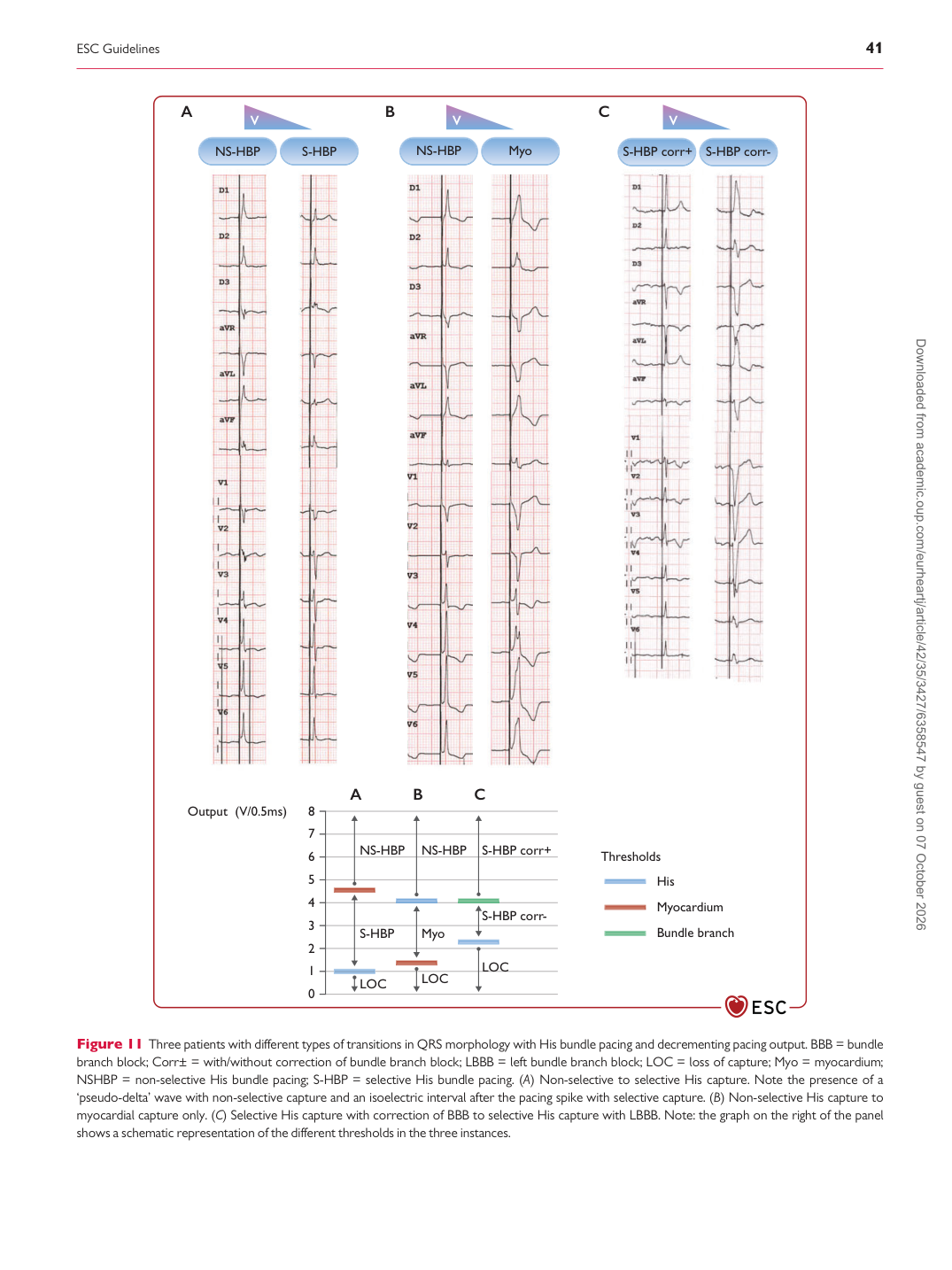
Figure 11 Three patients with different types of transitions in QRS morphology with His bundle pacing and decrementing pacing output. BBB = bundle branch block; Corr± = with/without correction of bundle branch block; LBBB = left bundle branch block; LOC = loss of capture; Myo = myocardium; NSHBP = non-selective His bundle pacing; S-HBP = selective His bundle pacing. (A) Non-selective to selective His capture. Note the presence of a ‘pseudo-delta’ wave with non-selective capture and an isoelectric interval after the pacing spike with selective capture. (B) Non-selective His capture to myocardial capture only. (C) Selective His capture with correction of BBB to selective His capture with LBBB. Note: the graph on the right of the panel shows a schematic representation of the different thresholds in the three instances. Downloaded from academic.oup.com/eurheartj/article/42/35/3427/6358547 by guest on 07 October 2026