p.47 抽出テキスト
................................................... • The evidence supporting the efficacy and safety of NOACs for the treatment of PE in patients with cancer needs to be extended by further studies. • In patients with cancer, the anticoagulant regimen and dose after the first 6 months should be clarified and prospectively tested. • The optimal time for discontinuing anticoagulant treatment after an episode of acute PE in patients with cancer is yet to be determined.

Pulmonary embolism and pregnancy • Diagnostic algorithms for PE in pregnancy, using modern radiological imaging techniques and low radiation doses, need to be prospectively tested in adequately powered cohort studies. • Controversy persists on the optimal LMWH dose and regimen for the treatment of PE during pregnancy. • NOACs are not allowed in pregnancy. However, if exposure to these drugs occurs during pregnancy despite this warning, any possible effects on the foetus should be recorded to provide more precise information on the risks and complications of these drugs, and adapt the instructions to physicians in the future.

Long-term sequelae of pulmonary embolism • The optimal follow-up strategy, including the spectrum of diagnostic tests that may be necessary, in patients with persisting symptoms and functional limitation after acute PE needs to be defined and prospectively validated. • In the absence of persisting symptoms or functional limitation after acute PE, the criteria for identifying patients whose risk of developing CTEPH may be sufficiently high to justify further diagnostic workup require further elaboration and validation in prospective cohort studies.

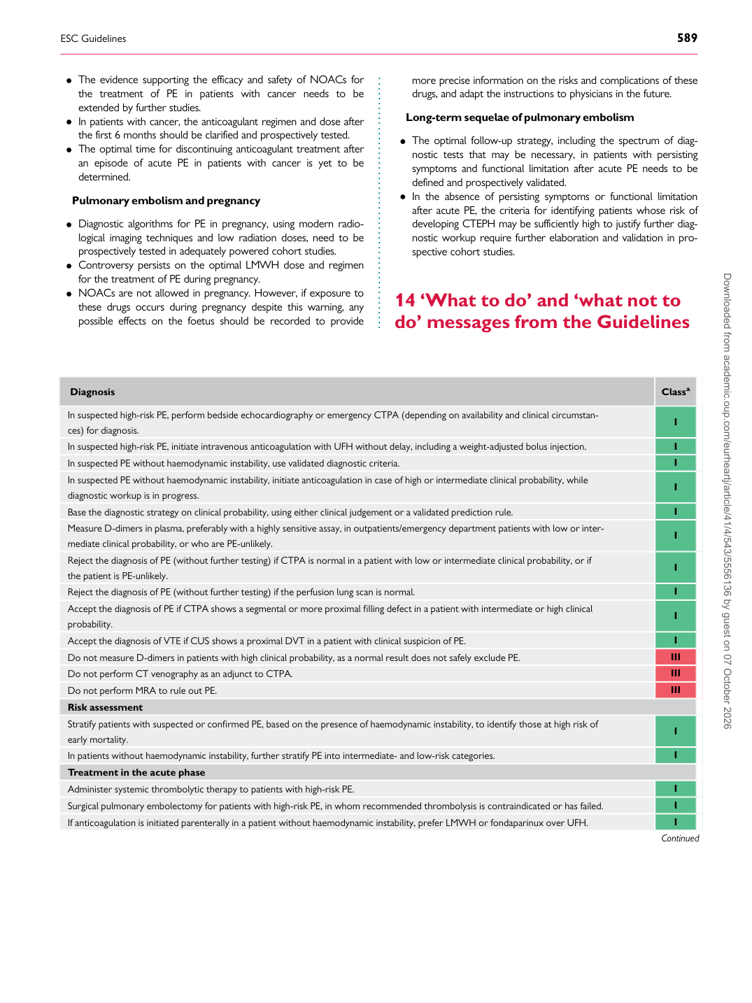
14 ‘What to do’ and ‘what not to do’ messages from the Guidelines

Diagnosis Classa

In suspected high-risk PE, perform bedside echocardiography or emergency CTPA (depending on availability and clinical circumstances) for diagnosis. I In suspected high-risk PE, initiate intravenous anticoagulation with UFH without delay, including a weight-adjusted bolus injection. I In suspected PE without haemodynamic instability, use validated diagnostic criteria. I In suspected PE without haemodynamic instability, initiate anticoagulation in case of high or intermediate clinical probability, while diagnostic workup is in progress. I Base the diagnostic strategy on clinical probability, using either clinical judgement or a validated prediction rule. I Measure D-dimers in plasma, preferably with a highly sensitive assay, in outpatients/emergency department patients with low or intermediate clinical probability, or who are PE-unlikely. I Reject the diagnosis of PE (without further testing) if CTPA is normal in a patient with low or intermediate clinical probability, or if the patient is PE-unlikely. I Reject the diagnosis of PE (without further testing) if the perfusion lung scan is normal. I Accept the diagnosis of PE if CTPA shows a segmental or more proximal filling defect in a patient with intermediate or high clinical probability. I Accept the diagnosis of VTE if CUS shows a proximal DVT in a patient with clinical suspicion of PE. I Do not measure D-dimers in patients with high clinical probability, as a normal result does not safely exclude PE. III Do not perform CT venography as an adjunct to CTPA. III Do not perform MRA to rule out PE. III Risk assessment Stratify patients with suspected or confirmed PE, based on the presence of haemodynamic instability, to identify those at high risk of early mortality. I In patients without haemodynamic instability, further stratify PE into intermediate- and low-risk categories. I Treatment in the acute phase Administer systemic thrombolytic therapy to patients with high-risk PE. I Surgical pulmonary embolectomy for patients with high-risk PE, in whom recommended thrombolysis is contraindicated or has failed. I If anticoagulation is initiated parenterally in a patient without haemodynamic instability, prefer LMWH or fondaparinux over UFH. I

Continued Downloaded from academic.oup.com/eurheartj/article/41/4/543/5556136 by guest on 07 October 2026