p.29 抽出テキスト
............................................ The diagnostic strategy for suspected acute PE in pregnancy is discussed in section 9.

7.1.1 Suspected pulmonary embolism with haemodynamic instability The proposed strategy is shown in Figure 4. The clinical probability is usually high and the differential diagnosis includes cardiac tamponade, acute coronary syndrome, aortic dissection, acute valvular dysfunction, and hypovolaemia. The most useful initial test in this situation is bedside TTE, which will yield evidence of acute RV dysfunction if acute PE is the cause of the patient’s haemodynamic decompensation. In a highly unstable patient, echocardiographic evidence of RV dysfunction is sufficient to prompt immediate reperfusion without further testing. This decision may be strengthened by the (rare) visualization of right heart thrombi.155,157,321,322 Ancillary bedside imaging tests include TOE, which may allow direct visualization of thrombi in the pulmonary artery and its main branches, especially in patients with RV dysfunction. TOE should be cautiously performed in hypoxaemic patients. Moreover, bedside CUS can detect proximal DVT. As soon as the patient is stabilized using supportive treatment, final confirmation of the diagnosis by CT angiography should be sought.

For unstable patients admitted directly to the catheterization laboratory with suspected acute coronary syndrome, pulmonary angiography may be considered as a diagnostic procedure after the acute coronary syndrome has been excluded, provided that PE is a probable diagnostic alternative and particularly if percutaneous catheterdirected treatment is a therapeutic option.

©ESC 2019

Suspected PE in a patient without haemodynamic instabilitya

Assess clinical probability of PE

Clinical judgement or prediction ruleb

Low or intermediate clinical probability,

or PE unlikely

Positive Negative

No PE PE confirmedd No PE PE confirmedd

High clinical probability

or PE likely

D-dimer test

CTPA CTPA

No treatmentc Treatmentc No treatmentc

or investigate

furthere Treatmentc

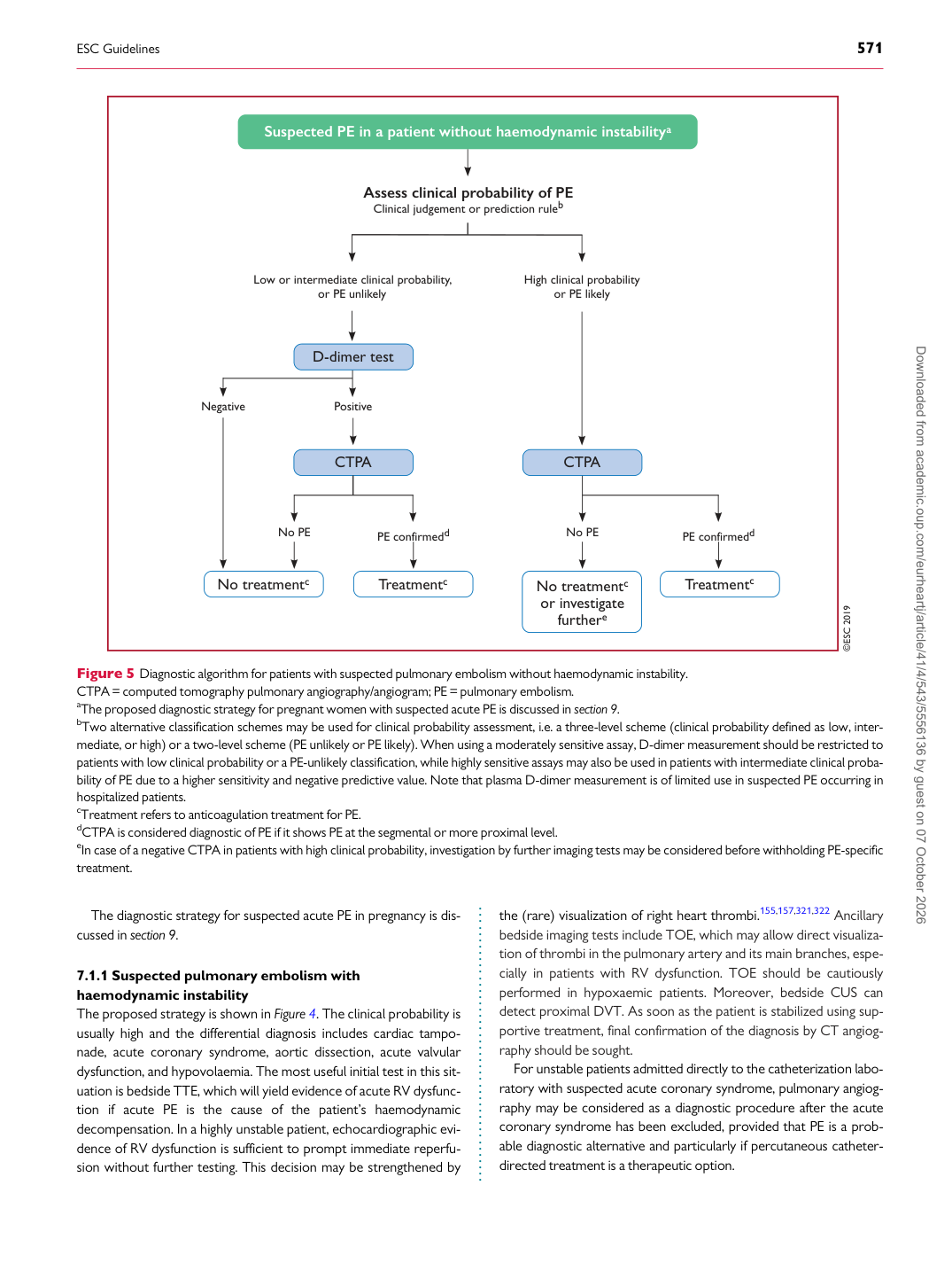
Figure 5 Diagnostic algorithm for patients with suspected pulmonary embolism without haemodynamic instability. CTPA= computed tomography pulmonary angiography/angiogram; PE = pulmonary embolism. aThe proposed diagnostic strategy for pregnant women with suspected acute PE is discussed in section 9. bTwo alternative classification schemes may be used for clinical probability assessment, i.e. a three-level scheme (clinical probability defined as low, intermediate, or high) or a two-level scheme (PE unlikely or PE likely). When using a moderately sensitive assay, D-dimer measurement should be restricted to patients with low clinical probability or a PE-unlikely classification, while highly sensitive assays may also be used in patients with intermediate clinical probability of PE due to a higher sensitivity and negative predictive value. Note that plasma D-dimer measurement is of limited use in suspected PE occurring in hospitalized patients. cTreatment refers to anticoagulation treatment for PE. dCTPA is considered diagnostic of PE if it shows PE at the segmental or more proximal level. eIn case of a negative CTPA in patients with high clinical probability, investigation by further imaging tests may be considered before withholding PE-specific treatment. Downloaded from academic.oup.com/eurheartj/article/41/4/543/5556136 by guest on 07 October 2026