p.28 抽出テキスト
...................................... lar retrieval of the permanent filter, or percutaneous nephrostomy or ureteral stent placement.306 Further reported complications include filter fracture and/or embolization, and DVT occasionally extending up to the vena cava.303,307,308

7 Integrated risk-adapted

diagnosis and management

7.1 Diagnostic strategies

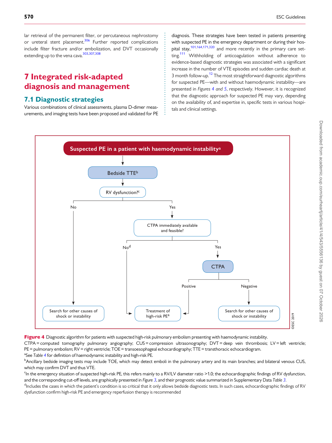
Various combinations of clinical assessments, plasma D-dimer measurements, and imaging tests have been proposed and validated for PE diagnosis. These strategies have been tested in patients presenting with suspected PE in the emergency department or during their hospital stay,101,164,171,320 and more recently in the primary care setting.111 Withholding of anticoagulation without adherence to evidence-based diagnostic strategies was associated with a significant increase in the number of VTE episodes and sudden cardiac death at 3 month follow-up.12 The most straightforward diagnostic algorithms for suspected PE—with and without haemodynamic instability—are presented in Figures 4 and 5, respectively. However, it is recognized that the diagnostic approach for suspected PE may vary, depending on the availability of, and expertise in, specific tests in various hospitals and clinical settings.

©ESC 2019

Bedside TTEb

CTPA

RV dysfunction?c

Treatment of

high-risk PEa Search for other causes of

shock or instability Search for other causes of

shock or instability

Suspected PE in a patient with haemodynamic instabilitya

Yes

Yes

Negative

No

Nod

Positive

CTPA immediately available

and feasible?

Figure 4 Diagnostic algorithm for patients with suspected high-risk pulmonary embolism presenting with haemodynamic instability. CTPA= computed tomography pulmonary angiography; CUS = compression ultrasonography; DVT = deep vein thrombosis; LV = left ventricle; PE= pulmonary embolism; RV = right ventricle; TOE= transoesophageal echocardiography; TTE = transthoracic echocardiogram. aSee Table 4 for definition of haemodynamic instability and high-risk PE. bAncillary bedside imaging tests may include TOE, which may detect emboli in the pulmonary artery and its main branches; and bilateral venous CUS, which may confirm DVT and thus VTE. cIn the emergency situation of suspected high-risk PE, this refers mainly to a RV/LV diameter ratio >1.0; the echocardiographic findings of RV dysfunction, and the corresponding cut-off levels, are graphically presented in Figure 3, and their prognostic value summarized in Supplementary Data Table 3. dIncludes the cases in which the patient’s condition is so critical that it only allows bedside diagnostic tests. In such cases, echocardiographic findings of RV dysfunction confirm high-risk PE and emergency reperfusion therapy is recommended Downloaded from academic.oup.com/eurheartj/article/41/4/543/5556136 by guest on 07 October 2026