p.9 抽出テキスト
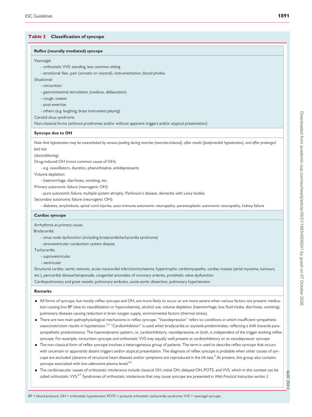
Table 3 Classification of syncope

Reflex (neurally mediated) syncope

Vasovagal:

- orthostatic VVS: standing, less common sitting - emotional: fear, pain (somatic or visceral), instrumentation, blood phobia Situational:

- micturition - gastrointestinal stimulation (swallow, defaecation) - cough, sneeze - post-exercise - others (e.g. laughing, brass instrument playing) Carotid sinus syndrome Non-classical forms (without prodromes and/or without apparent triggers and/or atypical presentation)

Syncope due to OH

Note that hypotension may be exacerbated by venous pooling during exercise (exercise-induced), after meals (postprandial hypotension), and after prolonged bed rest (deconditioning). Drug-induced OH (most common cause of OH):

- e.g. vasodilators, diuretics, phenothiazine, antidepressants Volume depletion: - haemorrhage, diarrhoea, vomiting, etc. Primary autonomic failure (neurogenic OH): - pure autonomic failure, multiple system atrophy, Parkinson’s disease, dementia with Lewy bodies Secondary autonomic failure (neurogenic OH):

- diabetes, amyloidosis, spinal cord injuries, auto-immune autonomic neuropathy, paraneoplastic autonomic neuropathy, kidney failure

Cardiac syncope

Arrhythmia as primary cause: Bradycardia:

- sinus node dysfunction (including bradycardia/tachycardia syndrome) - atrioventricular conduction system disease Tachycardia:

- supraventricular - ventricular Structural cardiac: aortic stenosis, acute myocardial infarction/ischaemia, hypertrophic cardiomyopathy, cardiac masses (atrial myxoma, tumours, etc.), pericardial disease/tamponade, congenital anomalies of coronary arteries, prosthetic valve dysfunction Cardiopulmonary and great vessels: pulmonary embolus, acute aortic dissection, pulmonary hypertension

Remarks • All forms of syncope, but mostly reflex syncope and OH, are more likely to occur or are more severe when various factors are present: medication causing low BP (due to vasodilatation or hypovolaemia), alcohol use, volume depletion (haemorrhage, low fluid intake, diarrhoea, vomiting), pulmonary diseases causing reduction in brain oxygen supply, environmental factors (thermal stress). • There are two main pathophysiological mechanisms in reflex syncope. “Vasodepression” refers to conditions in which insufficient sympathetic vasoconstriction results in hypotension.1,2 “Cardioinhibition” is used when bradycardia or asystole predominates, reflecting a shift towards para-

sympathetic predominance. The haemodynamic pattern, i.e. cardioinhibitory, vasodepressive, or both, is independent of the trigger evoking reflex syncope. For example, micturition syncope and orthostatic VVS may equally well present as cardioinhibitory or as vasodepressor syncope • The non-classical form of reflex syncope involves a heterogeneous group of patients. The term is used to describe reflex syncope that occurs

with uncertain or apparently absent triggers and/or atypical presentation. The diagnosis of reflex syncope is probable when other causes of syncope are excluded (absence of structural heart disease) and/or symptoms are reproduced in the tilt test.3 At present, this group also contains syncope associated with low adenosine plasma levels4,5 • The cardiovascular causes of orthostatic intolerance include classical OH, initial OH, delayed OH, POTS, and VVS, which in this context can be

called orthostatic VVS.6,7 Syndromes of orthostatic intolerance that may cause syncope are presented in Web Practical Instruction section 2.

BP = blood pressure; OH = orthostatic hypotension; POTS = postural orthostatic tachycardia syndrome; VVS = vasovagal syncope. Downloaded from academic.oup.com/eurheartj/article/39/21/1883/4939241 by guest on 07 October 2026