p.28 抽出テキスト
.................................................................... block could be the cause of unexplained syncope. Although cardiac pacing may lead to substantial reduction of syncopal attacks in elderly patients with unexplained syncope and a positive ATP test,229 previous studies showed no correlation between AV block induced by ATP and ECG findings (documented by ILR) during spontaneous syncope.122,123,227 Thus, the low predictive value of the test does not support its routine use in selecting patients for cardiac pacing, but rather its positivity suggests that it can be used to confirm the suspicion of asystolic syncope by means of prolonged ECG monitoring. The role of endogenous adenosine release in triggering a special form of asystolic syncope (so-called adenosine-sensitive syncope) remains under investigation.

4.2.7.2 Cardiovascular biomarkers Some cardiovascular biomarkers are increased in autonomic dysfunction underlying syncope, such as elevated copeptin (vasopressin), endothelin-1, and N-terminal pro-B-type natriuretic peptide in OH,113,230,231 whereas atrial natriuretic peptide may be reduced in POTS.113 At present, the use of cardiovascular biomarkers in the diagnosis of syncope awaits more evidence and verification in independent cohorts. 4.2.7.3 Immunological biomarkers Autoantibodies against adrenergic receptors in OH and POTS have been reported, but further studies are needed.232–234

4.2.8 Echocardiography For patients with suspected heart disease, echocardiography serves to confirm or refute the suspicions in equal proportions and plays an important role in risk stratification.235,236 Echocardiography identifies the cause of syncope in very few patients when no more tests are needed (i.e. severe aortic stenosis, obstructive cardiac tumours or thrombi, pericardial tamponade, or aortic dissection).237–239 In a literature review, right and left atrial myxoma presented with syncope in <20% of cases.240–244

4.2.8.1 Exercise stress echocardiography Upright or semi-supine exercise stress echocardiography to detect provocable left ventricular outflow tract obstruction should be considered in patients with HCM that complain of exertional or postural syncope, particularly when it recurs during similar circumstances (e.g. when rushing upstairs or straining). A gradient of >_50 mmHg is usually considered to be the threshold at which left ventricular outflow tract obstruction becomes haemodynamically important.245–249

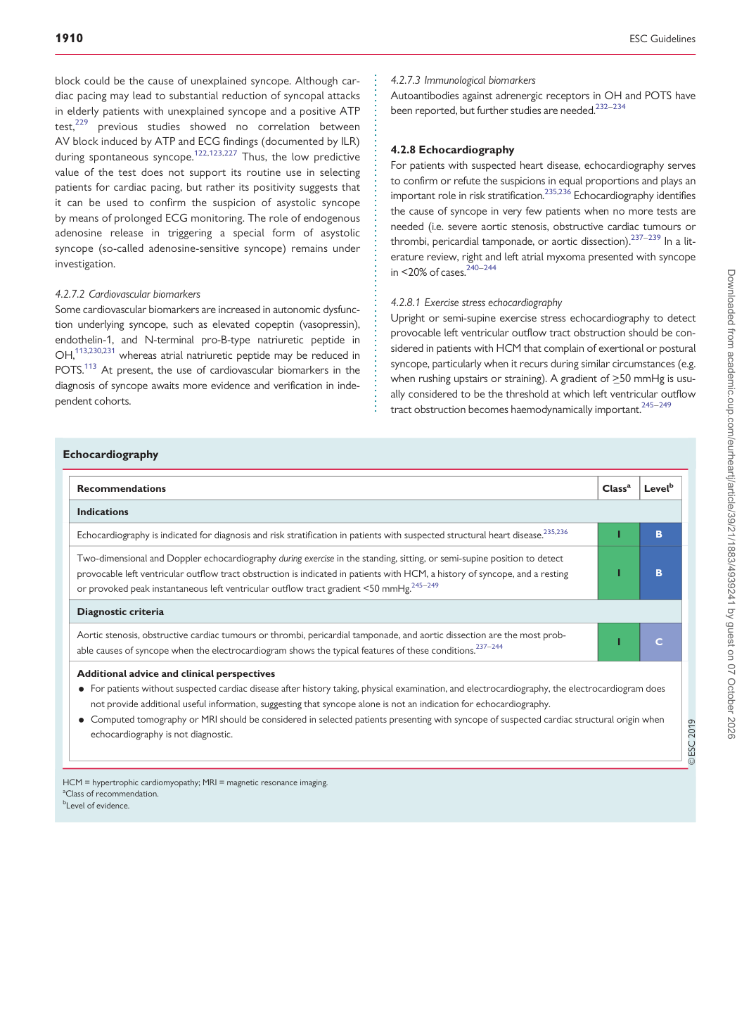
Echocardiography

Recommendations Classa Levelb

Indications Echocardiography is indicated for diagnosis and risk stratification in patients with suspected structural heart disease.235,236 I B

Two-dimensional and Doppler echocardiography during exercise in the standing, sitting, or semi-supine position to detect provocable left ventricular outflow tract obstruction is indicated in patients with HCM, a history of syncope, and a resting or provoked peak instantaneous left ventricular outflow tract gradient <50 mmHg.245–249 I B

Diagnostic criteria

Aortic stenosis, obstructive cardiac tumours or thrombi, pericardial tamponade, and aortic dissection are the most probable causes of syncope when the electrocardiogram shows the typical features of these conditions.237–244 I C

Additional advice and clinical perspectives • For patients without suspected cardiac disease after history taking, physical examination, and electrocardiography, the electrocardiogram does not provide additional useful information, suggesting that syncope alone is not an indication for echocardiography. • Computed tomography or MRI should be considered in selected patients presenting with syncope of suspected cardiac structural origin when echocardiography is not diagnostic.

HCM = hypertrophic cardiomyopathy; MRI = magnetic resonance imaging. aClass of recommendation. bLevel of evidence. Downloaded from academic.oup.com/eurheartj/article/39/21/1883/4939241 by guest on 07 October 2026