p.22 抽出テキスト
.............................................................................................................................................................................

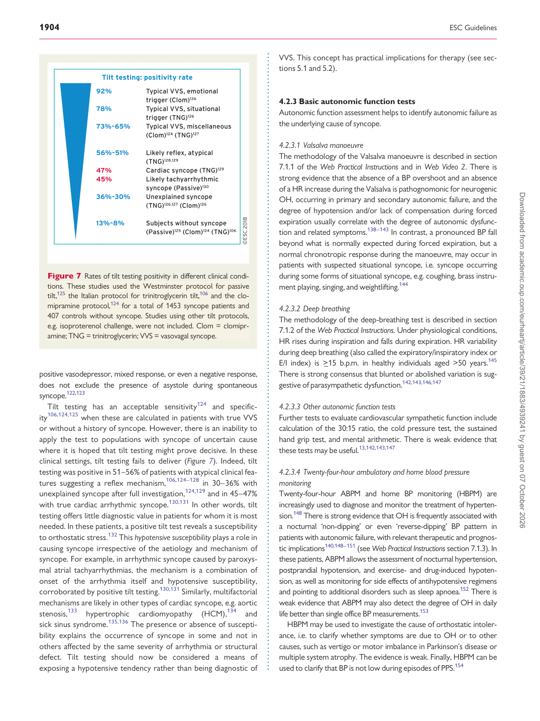
positive vasodepressor, mixed response, or even a negative response, does not exclude the presence of asystole during spontaneous syncope.122,123 Tilt testing has an acceptable sensitivity124 and specificity106,124,125 when these are calculated in patients with true VVS or without a history of syncope. However, there is an inability to apply the test to populations with syncope of uncertain cause where it is hoped that tilt testing might prove decisive. In these clinical settings, tilt testing fails to deliver (Figure 7). Indeed, tilt testing was positive in 51–56% of patients with atypical clinical features suggesting a reflex mechanism,106,124–128 in 30–36% with unexplained syncope after full investigation,124,129 and in 45–47% with true cardiac arrhythmic syncope.130,131 In other words, tilt testing offers little diagnostic value in patients for whom it is most needed. In these patients, a positive tilt test reveals a susceptibility to orthostatic stress.132 This hypotensive susceptibility plays a role in causing syncope irrespective of the aetiology and mechanism of syncope. For example, in arrhythmic syncope caused by paroxysmal atrial tachyarrhythmias, the mechanism is a combination of onset of the arrhythmia itself and hypotensive susceptibility, corroborated by positive tilt testing.130,131 Similarly, multifactorial mechanisms are likely in other types of cardiac syncope, e.g. aortic stenosis,133 hypertrophic cardiomyopathy (HCM),134 and sick sinus syndrome.135,136 The presence or absence of susceptibility explains the occurrence of syncope in some and not in others affected by the same severity of arrhythmia or structural defect. Tilt testing should now be considered a means of exposing a hypotensive tendency rather than being diagnostic of VVS. This concept has practical implications for therapy (see sections 5.1 and 5.2).

4.2.3 Basic autonomic function tests Autonomic function assessment helps to identify autonomic failure as the underlying cause of syncope.

4.2.3.1 Valsalva manoeuvre The methodology of the Valsalva manoeuvre is described in section 7.1.1 of the Web Practical Instructions and in Web Video 2. There is strong evidence that the absence of a BP overshoot and an absence of a HR increase during the Valsalva is pathognomonic for neurogenic OH, occurring in primary and secondary autonomic failure, and the degree of hypotension and/or lack of compensation during forced expiration usually correlate with the degree of autonomic dysfunction and related symptoms.138–143 In contrast, a pronounced BP fall beyond what is normally expected during forced expiration, but a normal chronotropic response during the manoeuvre, may occur in patients with suspected situational syncope, i.e. syncope occurring during some forms of situational syncope, e.g. coughing, brass instrument playing, singing, and weightlifting.144

4.2.3.2 Deep breathing The methodology of the deep-breathing test is described in section 7.1.2 of the Web Practical Instructions. Under physiological conditions, HR rises during inspiration and falls during expiration. HR variability during deep breathing (also called the expiratory/inspiratory index or E/I index) is >_15 b.p.m. in healthy individuals aged >50 years.145 There is strong consensus that blunted or abolished variation is suggestive of parasympathetic dysfunction.142,143,146,147

4.2.3.3 Other autonomic function tests Further tests to evaluate cardiovascular sympathetic function include calculation of the 30:15 ratio, the cold pressure test, the sustained hand grip test, and mental arithmetic. There is weak evidence that these tests may be useful.13,142,143,147

4.2.3.4 Twenty-four-hour ambulatory and home blood pressure monitoring Twenty-four-hour ABPM and home BP monitoring (HBPM) are increasingly used to diagnose and monitor the treatment of hypertension.148 There is strong evidence that OH is frequently associated with a nocturnal ‘non-dipping’ or even ‘reverse-dipping’ BP pattern in patients with autonomic failure, with relevant therapeutic and prognostic implications140,148–151 (see Web Practical Instructions section 7.1.3). In these patients, ABPM allows the assessment of nocturnal hypertension, postprandial hypotension, and exercise- and drug-induced hypotension, as well as monitoring for side effects of antihypotensive regimens and pointing to additional disorders such as sleep apnoea.152 There is weak evidence that ABPM may also detect the degree of OH in daily life better than single office BP measurements.153 HBPM may be used to investigate the cause of orthostatic intolerance, i.e. to clarify whether symptoms are due to OH or to other causes, such as vertigo or motor imbalance in Parkinson’s disease or multiple system atrophy. The evidence is weak. Finally, HBPM can be used to clarify that BP is not low during episodes of PPS.154

Figure 7 Rates of tilt testing positivity in different clinical conditions. These studies used the Westminster protocol for passive tilt,125 the Italian protocol for trinitroglycerin tilt,106 and the clomipramine protocol,124 for a total of 1453 syncope patients and 407 controls without syncope. Studies using other tilt protocols, e.g. isoproterenol challenge, were not included. Clom = clomipramine; TNG = trinitroglycerin; VVS = vasovagal syncope. Downloaded from academic.oup.com/eurheartj/article/39/21/1883/4939241 by guest on 07 October 2026