p.12 抽出テキスト
...................... • Carotid sinus massage (CSM) in patients aged >40 years. • Head-up tilt testing when there is suspicion of syncope due to OH or reflex syncope. • Blood tests when clinically indicated, e.g. haematocrit or haemoglobin when haemorrhage is suspected, oxygen saturation and blood gas analysis when hypoxia is suspected, troponin when cardiac ischaemia-related syncope is suspected, or D-dimer when pulmonary embolism is suspected, etc. Even if there is no independent gold/reference standard to diagnose syncope, there is strong consensus that the initial evaluation may lead to certain or highly likely diagnosis when the diagnostic criteria listed in the table of recommendations are met.

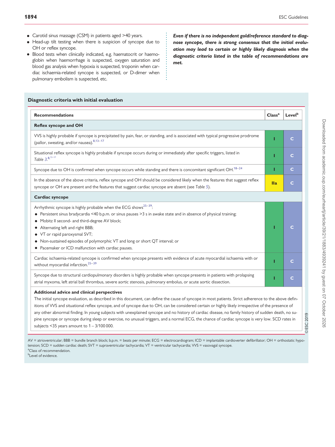
Diagnostic criteria with initial evaluation

Recommendations Classa Levelb

Reflex syncope and OH

VVS is highly probable if syncope is precipitated by pain, fear, or standing, and is associated with typical progressive prodrome (pallor, sweating, and/or nausea).8,13–17 I C

Situational reflex syncope is highly probable if syncope occurs during or immediately after specific triggers, listed in Table 3.8,13–17 I C Syncope due to OH is confirmed when syncope occurs while standing and there is concomitant significant OH.18–24 I C

In the absence of the above criteria, reflex syncope and OH should be considered likely when the features that suggest reflex syncope or OH are present and the features that suggest cardiac syncope are absent (see Table 5). IIa C

Cardiac syncope Arrhythmic syncope is highly probable when the ECG shows25–39: • Persistent sinus bradycardia <40 b.p.m. or sinus pauses >3 s in awake state and in absence of physical training; • Mobitz II second- and third-degree AV block; • Alternating left and right BBB; • VT or rapid paroxysmal SVT; • Non-sustained episodes of polymorphic VT and long or short QT interval; or • Pacemaker or ICD malfunction with cardiac pauses. I C Cardiac ischaemia-related syncope is confirmed when syncope presents with evidence of acute myocardial ischaemia with or without myocardial infarction.25–39 I C

Syncope due to structural cardiopulmonary disorders is highly probable when syncope presents in patients with prolapsing atrial myxoma, left atrial ball thrombus, severe aortic stenosis, pulmonary embolus, or acute aortic dissection. I C

Additional advice and clinical perspectives The initial syncope evaluation, as described in this document, can define the cause of syncope in most patients. Strict adherence to the above definitions of VVS and situational reflex syncope, and of syncope due to OH, can be considered certain or highly likely irrespective of the presence of any other abnormal finding. In young subjects with unexplained syncope and no history of cardiac disease, no family history of sudden death, no supine syncope or syncope during sleep or exercise, no unusual triggers, and a normal ECG, the chance of cardiac syncope is very low. SCD rates in subjects <35 years amount to 1 – 3/100 000.

AV = atrioventricular; BBB = bundle branch block; b.p.m. = beats per minute; ECG = electrocardiogram; ICD = implantable cardioverter defibrillator; OH = orthostatic hypotension; SCD = sudden cardiac death; SVT = supraventricular tachycardia; VT = ventricular tachycardia; VVS = vasovagal syncope. aClass of recommendation. bLevel of evidence. Downloaded from academic.oup.com/eurheartj/article/39/21/1883/4939241 by guest on 07 October 2026