p.43 抽出テキスト
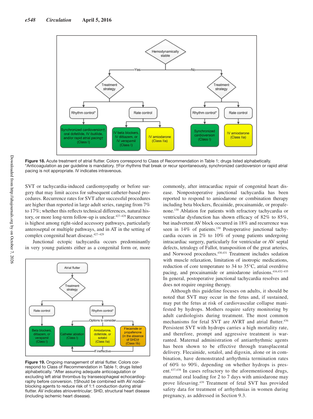
SVT or tachycardia-induced cardiomyopathy or before surgery that may limit access for subsequent catheter-based procedures. Recurrence rates for SVT after successful procedures are higher than reported in large adult series, ranging from 7% to 17%; whether this reflects technical differences, natural history, or more long-term follow-up is unclear.427–429 Recurrence is highest among right-sided accessory pathways, particularly anteroseptal or multiple pathways, and in AT in the setting of complex congenital heart disease.427–429 Junctional ectopic tachycardia occurs predominantly in very young patients either as a congenital form or, more commonly, after intracardiac repair of congenital heart disease. Nonpostoperative junctional tachycardia has been reported to respond to amiodarone or combination therapy including beta blockers, flecainide, procainamide, or propafenone.130 Ablation for patients with refractory tachycardia or ventricular dysfunction has shown efficacy of 82% to 85%, but inadvertent AV block occurred in 18% and recurrence was seen in 14% of patients.130 Postoperative junctional tachycardia occurs in 2% to 10% of young patients undergoing intracardiac surgery, particularly for ventricular or AV septal defects, tetralogy of Fallot, transposition of the great arteries, and Norwood procedures.430,431 Treatment includes sedation with muscle relaxation, limitation of inotropic medications, reduction of core temperature to 34 to 35°C, atrial overdrive pacing, and procainamide or amiodarone infusions.416,432–435 In general, postoperative junctional tachycardia resolves and does not require ongoing therapy. Although this guideline focuses on adults, it should be noted that SVT may occur in the fetus and, if sustained, may put the fetus at risk of cardiovascular collapse manifested by hydrops. Mothers require safety monitoring by adult cardiologists during treatment. The most common mechanisms for fetal SVT are AVRT and atrial flutter.436 Persistent SVT with hydrops carries a high mortality rate, and therefore, prompt and aggressive treatment is warranted. Maternal administration of antiarrhythmic agents has been shown to be effective through transplacental delivery. Flecainide, sotalol, and digoxin, alone or in combination, have demonstrated arrhythmia termination rates of 60% to 90%, depending on whether hydrops is present.437,438 In cases refractory to the aforementioned drugs, maternal oral loading for 2 to 7 days with amiodarone may prove lifesaving.439 Treatment of fetal SVT has provided safety data for treatment of arrhythmias in women during pregnancy, as addressed in Section 9.3. Figure 18. Acute treatment of atrial flutter. Colors correspond to Class of Recommendation in Table 1; drugs listed alphabetically. *Anticoagulation as per guideline is mandatory. †For rhythms that break or recur spontaneously, synchronized cardioversion or rapid atrial pacing is not appropriate. IV indicates intravenous. Figure 19. Ongoing management of atrial flutter. Colors correspond to Class of Recommendation in Table 1; drugs listed alphabetically. *After assuring adequate anticoagulation or excluding left atrial thrombus by transesophageal echocardiography before conversion. †Should be combined with AV nodal– blocking agents to reduce risk of 1:1 conduction during atrial flutter. AV indicates atrioventricular; SHD, structural heart disease (including ischemic heart disease). Downloaded from http://ahajournals.org by on October 7, 2026