p.35 抽出テキスト
recommendations for anticoagulation mirror those for patients with AF.10,121,314 Similarly, the recommendations for anticoagulation with regard to either pharmacological or electrical cardioversion of patients with atrial flutter are the same as those for patients with AF, as discussed in the 2014 AF guideline (Section 6.1).10

7.2 Non–Isthmus-Dependent Atrial Flutters

Non–isthmus-dependent atrial flutter or atypical flutter describes macroreentrant ATs that are not dependent on conduction through the CTI. A variety of circuits have been described, including a path around the mitral annulus (perimitral flutter), re-entry involving the left atrial roof, and re-entry around regions of scarring in the right or left atrium. Non– isthmus-dependent atrial flutters often occur in patients with atrial scarring from prior heart surgery or ablation but also may occur in any form of heart disease or may be idiopathic.134,140,319 Non–isthmus-dependent atrial flutters can coexist with CTIdependent flutter or involve the presence of multiple atrial reentry circuits.133,320 The reentrant circuits are classified as either macroreentrant AT (large; often several centimeters or longer in diameter) or microreentrant AT (≤2 cm in diameter), which may be indistinguishable from focal AT.321 In the presence of substantial atrial disease, prior surgery, or prior radiofrequency catheter ablation, the ECG flutterwave morphology is not a reliable predictor of whether the flutter circuit involves the CTI. Although an ECG with a typical flutter appearance has good predictive value for CTIdependent flutter in a patient who has not undergone prior catheter ablation of AF, the ECG appearance is less useful in predicting the flutter circuit in a patient who has previously undergone AF ablation.322–325 The presence of a positive or biphasic (but dominantly positive) deflection in V1, accompanied by deflections in other leads inconsistent with typical counterclockwise atrial flutter, suggests the presence of an atypical flutter (Figure 17). Definitive diagnosis requires EP study and intracardiac mapping.326

Catheter ablation of non–CTI-dependent flutter requires more extensive mapping than does ablation of CTI-dependent flutter, and success rates are lower (Table 8). The location of the circuit determines ablation approach and risks. The substrate for macroreentrant atrial arrhythmias after cardiac surgery is atrial scarring from atriotomy incisions and cannulation sites and from the underlying myopathic process of the valve disease itself; this is sometimes referred to as incisional atrial reentrant tachycardia. The location of the reentrant circuit depends on the type of surgical approach, and common populations include patients who have undergone mitral valve surgery or have a repaired atrial septal defect.327–330 These arrhythmias are also common after surgical or catheter ablation for AF.331,332 Both single- and dual-loop circuits, as well as focal ATs, can be present. It is useful to review the procedural notes to identify the location of atrial incisions or prior ablation that can assist with future mapping and ablation. The development of a microreentrant or macroreentrant left AT after AF ablation occurs in approximately 5% of patients.330,333,334 This is less frequent if ablation is limited to pulmonary vein isolation. On the other hand, these arrhythmias

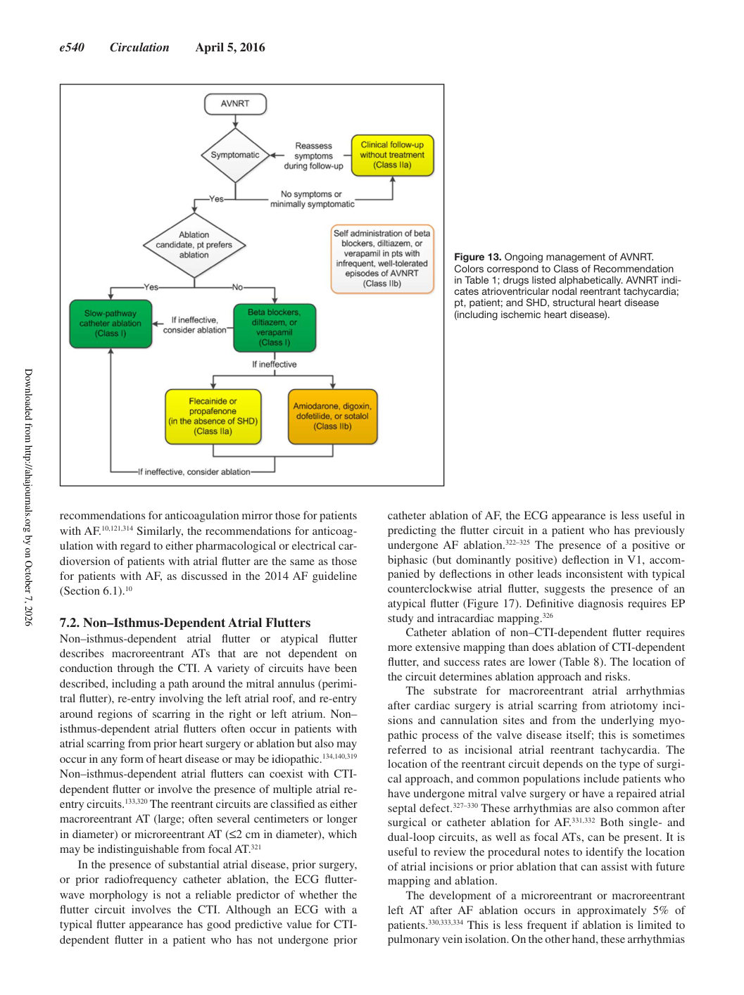
Figure 13. Ongoing management of AVNRT. Colors correspond to Class of Recommendation in Table 1; drugs listed alphabetically. AVNRT indicates atrioventricular nodal reentrant tachycardia; pt, patient; and SHD, structural heart disease (including ischemic heart disease).

Downloaded from http://ahajournals.org by on October 7, 2026