p.14 抽出テキスト
success rates comparable to conventional approaches yet with significant reduction in fluoroscopy times.145–148

2.5.3 Mapping and Ablation With No or Minimal Radiation

Fluoroscopy has historically been the primary imaging modality used for EP studies. The use of ionizing radiation puts patient, operator, and laboratory staff at risk of the short- and long-term effects of radiation exposure. Attention to optimal fluoroscopic technique and adoption of radiation-reducing strategies can minimize radiation dose to the patient and operator. The current standard is to use the “as low as reasonably achievable” (ALARA) principle on the assumption that there is no threshold below which ionizing radiation is free from harmful biological effect. Alternative imaging systems, such as electroanatomic mapping and intracardiac echocardiography, have led to the ability to perform SVT ablation with no or minimal fluoroscopy, with success and complication rates similar to standard techniques.147,149–152 Radiation exposure may be further reduced by using robotic or magnetic navigation of catheters that use a 3-dimensional anatomic tracking system superimposed on traditional fluoroscopy imaging. A reduced- fluoroscopy approach is particularly important in pediatric patients and during pregnancy.153,154

2.5.4 Ablation Energy Sources

Radiofrequency current is the most commonly used energy source for SVT ablation.155 Cryoablation is used as an alternative to radiofrequency ablation to minimize injury to the AV node during ablation of specific arrhythmias, such as AVNRT, para-Hisian AT, and para-Hisian accessory pathways, particularly in specific patient populations, such as children and young adults. Selection of the energy source

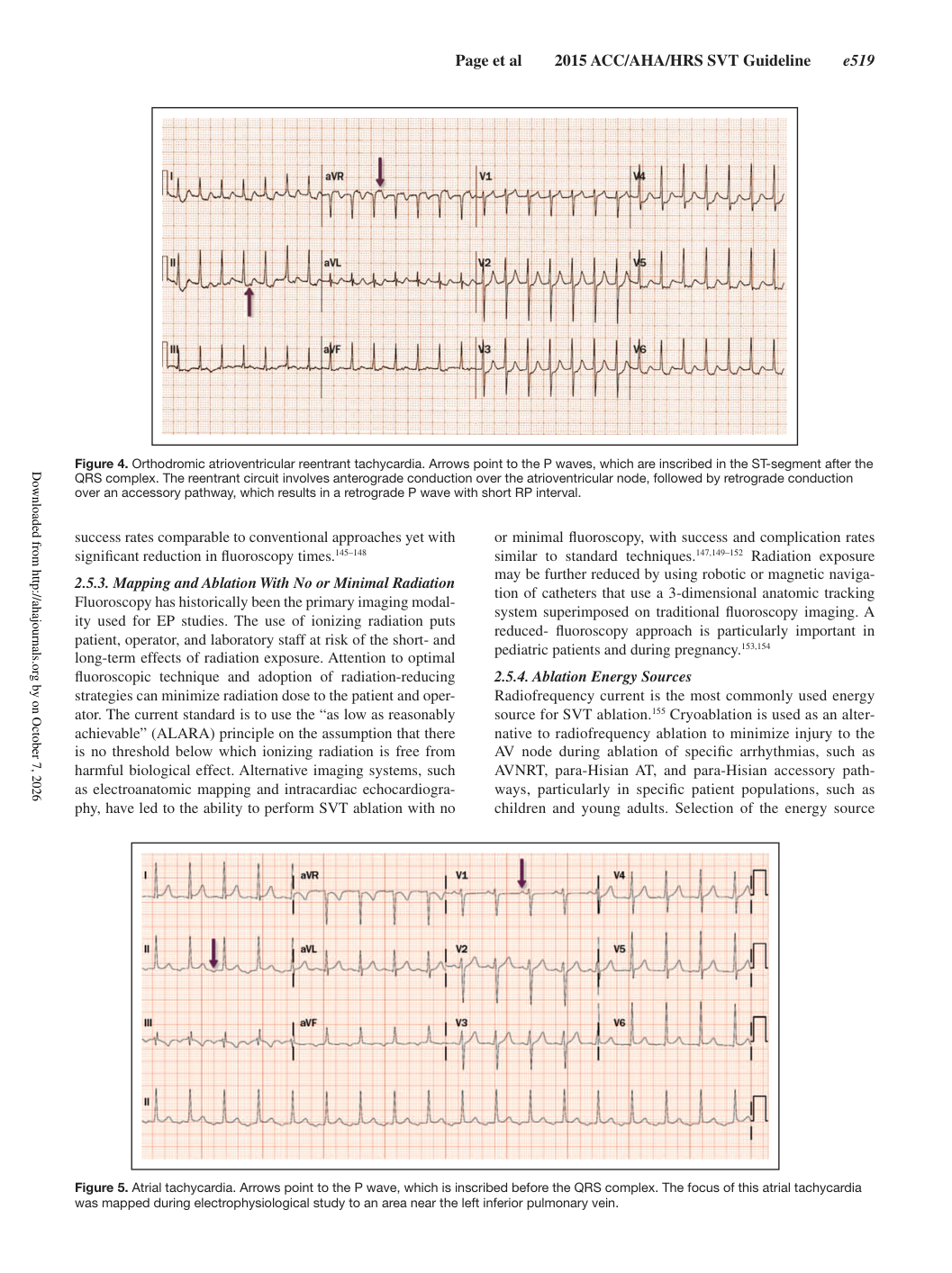
Figure 4. Orthodromic atrioventricular reentrant tachycardia. Arrows point to the P waves, which are inscribed in the ST-segment after the QRS complex. The reentrant circuit involves anterograde conduction over the atrioventricular node, followed by retrograde conduction over an accessory pathway, which results in a retrograde P wave with short RP interval.

Figure 5. Atrial tachycardia. Arrows point to the P wave, which is inscribed before the QRS complex. The focus of this atrial tachycardia was mapped during electrophysiological study to an area near the left inferior pulmonary vein.

Downloaded from http://ahajournals.org by on October 7, 2026